
Hydrocoele, Varicocoele and Epididymal Cyst
Key points
- The three questions: Can you get above it? (no = inguinoscrotal hernia). Does it transilluminate? (yes = fluid). Is it separate from the testis? (yes = epididymal, and usually benign).
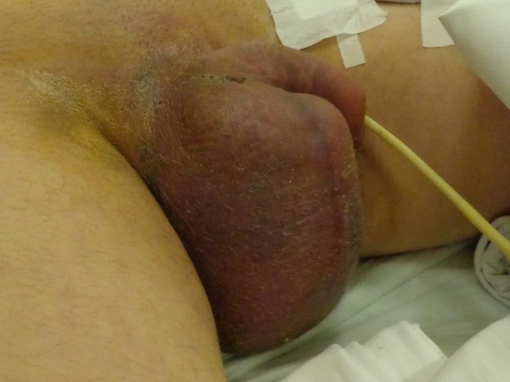
- Hydrocoele: fluid within the tunica vaginalis. A smooth, fluctuant, non-tender swelling that transilluminates, in which the testis is impalpable because it lies within the fluid.
- Varicocoele: dilated pampiniform plexus veins - a 'bag of worms' that is more prominent on standing and decompresses on lying flat. 80-90% are left-sided.
- Epididymal cyst / spermatocoele: a smooth cystic swelling separate from and above/behind the testis, which transilluminates. A spermatocoele contains sperm; both are benign.
- Red flag 1 - the hidden tumour: any hydrocoele in a young man, or a new-onset hydrocoele, needs an ultrasound - a secondary hydrocoele can conceal a testicular tumour.
- Red flag 2 - the renal tumour: a varicocoele that does not empty on lying flat, is right-sided, or is of sudden onset in an older man, suggests renal cell carcinoma obstructing the left renal vein.
- Fertility: varicocoele is the commonest correctable cause of male subfertility, associated with impaired sperm count and motility from raised scrotal temperature.
- Management: mostly conservative with reassurance. Intervene for symptoms, cosmetic concern, subfertility, or testicular growth arrest in adolescents.
Introduction
Scrotal swellings are common and the great majority are benign. What matters clinically is a systematic approach that reliably separates the benign from the serious - specifically from testicular tumour, inguinoscrotal hernia, and the small group of presentations that signal disease elsewhere.1
| Feature | Hydrocoele | Varicocoele | Epididymal cyst | Testicular tumour | Inguinoscrotal hernia |
|---|---|---|---|---|---|
| Get above it? | Yes | Yes | Yes | Yes | No |
| Transilluminates? | Yes | No | Yes | No | No |
| Relation to testis | Surrounds it - testis impalpable | Above and behind, along the cord | Separate, above/behind testis | Arises from the testis | Separate, extends to inguinal canal |
| Consistency | Smooth, fluctuant | 'Bag of worms' | Smooth, cystic | Hard, irregular | Soft, may be reducible |
| Effect of lying flat | No change | Decompresses | No change | No change | Often reduces |
| Tenderness | Usually none | Usually none (may ache) | None | Usually painless | Tender if strangulated |
| Cough impulse | No | May be present (Valsalva) | No | No | Yes |
Hydrocoele
A hydrocoele is an abnormal collection of serous fluid within the tunica vaginalis, the potential space surrounding the testis.
Classification and causes
- Communicating (congenital) hydrocoele - the processus vaginalis fails to obliterate, allowing peritoneal fluid to track into the scrotum. Common in infants, characteristically varies in size through the day (larger by evening), and the great majority resolve spontaneously by 1-2 years. Because the same patency underlies indirect inguinal hernia, the two are related
- Non-communicating (primary) hydrocoele - the commonest type in adults; idiopathic, from imbalance between fluid secretion and absorption by the tunica. Typically middle-aged or older men, gradual and painless
- Secondary hydrocoele - reactive to underlying pathology, and the type that must not be missed: testicular tumour, epididymo-orchitis, torsion, trauma, or rarely filariasis (a major cause worldwide but rare in the UK)
Clinical features
- Painless, smooth, fluctuant scrotal swelling that may become large and heavy
- Transilluminates brightly
- You can get above it
- The testis is typically impalpable within the fluid - the key limitation of clinical examination
- Non-tender, unless secondary to infection or torsion
- A rapidly developing or painful hydrocoele, or one in a young man, should raise concern about an underlying cause
Management
- Ultrasound - to confirm the diagnosis and, crucially, to visualise the underlying testis and exclude a tumour. Essential in young men, in new or rapidly enlarging hydrocoeles, and whenever the testis is impalpable
- Infants - conservative management with reassurance; most resolve by 1-2 years. Surgical repair (ligation of the patent processus vaginalis) if it persists beyond 2 years or if a hernia is present
- Adults - conservative management is appropriate for small, asymptomatic hydrocoeles once a tumour has been excluded
- Surgery - for discomfort, large size, cosmetic concerns or interference with daily activities. Options are Jaboulay (eversion of the sac) or Lord's plication procedures
- Aspiration with or without sclerotherapy - reserved for men unfit for surgery, as recurrence is common and there is an infection risk. Never aspirate before excluding a tumour
Varicocoele
A varicocoele is abnormal dilatation and tortuosity of the pampiniform plexus of veins draining the testis, caused by incompetent or absent venous valves and consequent venous reflux. It affects around 15% of men and up to 40% of men presenting with infertility.
Clinical features
- Often asymptomatic, found incidentally or during fertility investigation
- A dragging, aching or heavy sensation in the scrotum, worse on standing, at the end of the day, or after exertion
- 'Bag of worms' texture on palpation, above and behind the testis
- More prominent on standing and on Valsalva; decompresses on lying flat - the cardinal feature
- Testicular atrophy on the affected side, particularly relevant in adolescents
- Subfertility - impaired sperm count, motility and morphology
- Graded clinically: grade 1 palpable only on Valsalva; grade 2 palpable at rest; grade 3 visible through the scrotal skin
Investigation and management
- Scrotal ultrasound with Doppler - confirms venous dilatation (typically veins over 3 mm with reflux on Valsalva) and allows measurement of testicular volume
- CT abdomen - if the varicocoele is right-sided, of sudden onset, or fails to decompress
- Semen analysis - in men with fertility concerns
- Conservative management with reassurance and scrotal support is appropriate for most - the majority are asymptomatic and need no treatment
- Indications for intervention: persistent pain, testicular atrophy or growth arrest in adolescents, subfertility with abnormal semen parameters, or significant cosmetic concern
- Treatment options: surgical ligation (varicocoelectomy) - open, laparoscopic or microsurgical subinguinal (which has the lowest recurrence and complication rates) - or radiological percutaneous embolisation
- Complications of treatment: recurrence, hydrocoele formation (from lymphatic disruption), and rarely testicular atrophy
- Repair improves semen parameters in appropriately selected men, though the effect on live birth rates remains debated
Epididymal cyst and spermatocoele
Epididymal cysts are benign fluid-filled swellings arising from the epididymis, most often at its head. A spermatocoele is essentially the same lesion but contains sperm, giving milky rather than clear fluid; the distinction is made on aspiration or histology and has no practical significance.
Clinical features
- Smooth, round, fluctuant swelling, typically at the head of the epididymis
- Distinctly separate from and lying above and behind the testis - the key discriminating sign, since a normal testis can be palpated separately
- Transilluminates
- Painless, though larger cysts may cause a dragging sensation
- Often multiple and bilateral
- Common, increasingly so with age, and found in a substantial proportion of men on ultrasound
Management
- Ultrasound to confirm the diagnosis where there is any doubt about its relationship to the testis
- Conservative management with reassurance is appropriate for the great majority - these are benign and do not become malignant
- Surgical excision only for significant pain, very large size or cosmetic concern
- Counsel men of reproductive age before surgery: excision carries a risk of epididymal obstruction and consequent impairment of fertility on that side, so intervention should not be undertaken lightly in a man who may wish to father children
- Aspiration is not generally recommended because of high recurrence rates
Other scrotal swellings to recognise
- Inguinoscrotal hernia - you cannot get above it; there may be a cough impulse, reducibility and bowel sounds. Irreducible, tense and tender with vomiting suggests strangulation - a surgical emergency
- Testicular tumour - hard, irregular, arising from the testis, does not transilluminate. Urgent referral and ultrasound
- Testicular torsion - sudden severe pain, high-riding horizontal testis, absent cremasteric reflex; emergency exploration
- Epididymo-orchitis - painful, tender, warm, often febrile with urinary or sexual symptoms; positive Prehn's sign
- Haematocoele - blood within the tunica vaginalis after trauma; does not transilluminate, and testicular rupture must be excluded
- Sebaceous cysts of the scrotal skin - clearly within the skin and mobile over the underlying contents
- Idiopathic scrotal oedema - in children, painless diffuse swelling and erythema
- Scrotal or testicular abscess - fluctuant, tender, febrile
Red flags
Prognosis
All three of these conditions are benign, and the prognosis is excellent. None undergoes malignant change, and the majority of patients need nothing more than an accurate diagnosis and clear reassurance. Congenital hydrocoeles resolve spontaneously in the great majority of infants by the age of 1 to 2 years, and adult hydrocoeles and epididymal cysts can be safely observed indefinitely provided an underlying tumour has been excluded.
Surgical outcomes are generally good but not without cost. Hydrocoele repair is effective, with recurrence rates well below those of aspiration, which recurs commonly and is therefore reserved for men unfit for surgery. Excision of epididymal cysts carries a genuine risk of epididymal obstruction and impaired fertility on that side, which is why conservative management is strongly preferred in men who may wish to have children - a point that is easy to overlook when a patient simply wants a lump removed.
Varicocoele has the most nuanced prognosis because of its fertility implications. It is the commonest surgically correctable cause of male subfertility, and repair reliably improves semen parameters in appropriately selected men with abnormal analyses. The evidence that this translates into higher live birth rates is less consistent, so counselling should be honest about that uncertainty rather than promising a specific outcome. In adolescents, the important consideration is testicular growth: a varicocoele associated with reduced testicular volume on the affected side warrants referral, since timely repair can allow catch-up growth. Finally, the durable message across all three conditions is that the clinical value lies less in treating them than in confidently excluding the serious diagnoses they can mimic or conceal - testicular tumour beneath a hydrocoele, and renal cell carcinoma behind a non-decompressing varicocoele.
References
- NICE Clinical Knowledge Summaries. Scrotal pain and swelling. Available here
- European Association of Urology. Guidelines on Sexual and Reproductive Health - varicocoele and male infertility. Available here
- British Association of Urological Surgeons (BAUS). Hydrocele, epididymal cyst and varicocele - patient information. Available here
- NICE NG12. Suspected cancer: recognition and referral. 2015, updated 2023. Available here
- Jungwirth A, Diemer T, Kopa Z et al. EAU Guidelines on Male Infertility. Available here
- European Association of Urology. Guidelines on Paediatric Urology - hydrocele and varicocele in children. Available here
- AfroBrazilian, CC BY-SA 3.0, via Wikimedia Commons. Available here
This article is written for revision and education. It is not clinical guidance and must not be used to make decisions about the care of a patient. Always check current NICE guidance and local protocols.