
Polycystic Kidney Disease: Genetics, Extrarenal Disease and Tolvaptan
Key points
- Autosomal dominant PKD (ADPKD): the commonest inherited kidney disease, affecting around 1 in 1,000 people, in which progressive bilateral cyst formation destroys renal parenchyma over decades.
- Two genes: PKD1 on chromosome 16 (around 85% of cases, encoding polycystin-1) causes earlier renal failure - median around 55 years. PKD2 on chromosome 4 is milder, with failure around 70.
- Presentation: hypertension in a young adult, loin or abdominal pain, haematuria, recurrent urinary infection, palpable kidneys, or an incidental finding while screening a family member.
- Extrarenal disease is the key exam material: hepatic cysts (commonest), intracranial berry aneurysms with risk of subarachnoid haemorrhage, mitral valve prolapse, colonic diverticula and abdominal wall hernias.
- Diagnosis: ultrasound with age-adjusted cyst criteria in a patient with a positive family history; genetic testing where imaging is equivocal or for reproductive planning.
- Disease-modifying treatment: tolvaptan, a vasopressin V2 receptor antagonist, slows cyst growth and eGFR decline in rapidly progressing disease. It causes marked thirst, polyuria and hepatotoxicity needing LFT monitoring.
- Supportive management: rigorous blood pressure control with an ACE inhibitor or ARB, high fluid intake, low sodium diet, and prompt treatment of infection and stones.
- ARPKD: autosomal recessive PKD presents in infancy with enlarged echogenic kidneys, Potter sequence and pulmonary hypoplasia, plus congenital hepatic fibrosis - a distinct and far more severe disease.
Introduction
Polycystic kidney disease is a group of inherited disorders characterised by the development of multiple fluid-filled cysts that progressively replace functioning renal parenchyma. Autosomal dominant polycystic kidney disease (ADPKD) is by far the commoner form, affecting around 1 in 1,000 people, making it the commonest inherited kidney disease and the fourth commonest cause of end-stage renal disease in the UK.1
It is fundamentally a systemic disorder rather than a purely renal one. The polycystin proteins are expressed in epithelial cilia throughout the body, which is why cysts also form in the liver and pancreas, and why the vasculature and connective tissues are affected - producing the intracranial aneurysms, valvular disease and hernias that make up much of the clinically important burden.
| Feature | ADPKD | ARPKD |
|---|---|---|
| Inheritance | Autosomal dominant - 50% risk to each offspring; there is usually a family history, though around 10% arise de novo | Autosomal recessive - 25% risk with two carrier parents; parents are unaffected |
| Gene | PKD1 (chromosome 16, ~85%) or PKD2 (chromosome 4, ~15%) | PKHD1 (chromosome 6), encoding fibrocystin |
| Age at presentation | Adulthood, typically third to fifth decade | Neonatal or antenatal |
| Kidneys | Progressively enlarging cysts of varying size, massively enlarged in advanced disease | Bilaterally enlarged, diffusely echogenic kidneys with microcysts |
| Liver | Hepatic cysts (very common, rarely cause dysfunction) | Congenital hepatic fibrosis with portal hypertension |
| Prognosis | End-stage renal disease typically in the fifth to seventh decade | High perinatal mortality from pulmonary hypoplasia; survivors usually reach ESRD in childhood or adolescence |
Genetics and pathophysiology
| Gene | Chromosome | Protein | Proportion | Severity |
|---|---|---|---|---|
| PKD1 | 16 | Polycystin-1 | ~85% | More severe - larger kidneys, earlier hypertension, median age at end-stage renal disease around 55 years |
| PKD2 | 4 | Polycystin-2 | ~15% | Milder - later presentation, median age at end-stage renal disease around 70 years, and some never reach it |
Polycystin-1 and polycystin-2 form a complex in the primary cilium of renal tubular epithelial cells, where they act as a mechanosensor detecting urinary flow and regulating intracellular calcium. Loss of function reduces intracellular calcium and raises cyclic AMP, which drives epithelial cell proliferation and fluid secretion into the tubular lumen. Segments of tubule progressively balloon into fluid-filled cysts that detach from the nephron and continue to enlarge.
- Cysts arise from only a small proportion of nephrons, but their relentless expansion compresses and destroys the surrounding normal parenchyma, causing interstitial fibrosis - which is why renal function declines even though most nephrons are not cystic
- Vasopressin drives the process by raising cyclic AMP through the V2 receptor. This is the mechanistic basis both for advising a high fluid intake (suppressing endogenous vasopressin) and for treating with tolvaptan (a V2 receptor antagonist).
- GFR is preserved for decades by compensatory hyperfiltration of surviving nephrons, so a normal creatinine in a young adult with ADPKD is entirely expected and does not indicate mild disease. Total kidney volume rises long before eGFR falls, and is the better early marker of progression.
- Hypertension develops early, often before any fall in eGFR, because cyst expansion causes local ischaemia and activates the renin-angiotensin-aldosterone system - and it then accelerates the decline in function
Clinical features
Renal manifestations
- Hypertension - present in the majority by early adulthood and frequently the first sign; ADPKD should be considered in any young person with unexplained hypertension and a relevant family history
- Loin, flank or abdominal pain - either chronic and dragging, from the mass effect of enlarging kidneys, or acute, from cyst haemorrhage, cyst infection or an obstructing stone
- Haematuria - visible or non-visible, commonly from cyst haemorrhage, which is usually self-limiting
- Recurrent urinary tract infection and cyst infection - cysts are poorly perfused and antibiotic penetration is limited
- Renal calculi - in around 20%, from urinary stasis, hypocitraturia and low urine pH
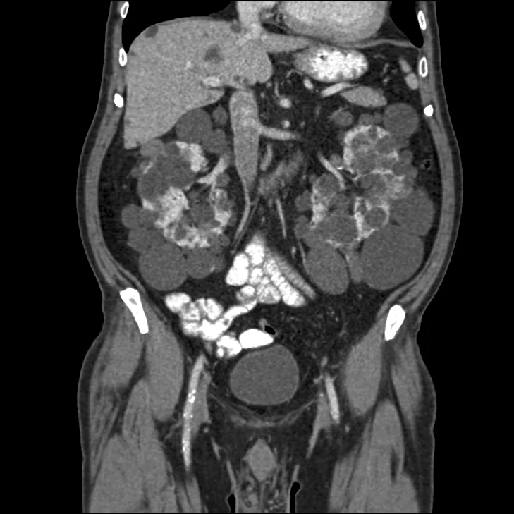
- Palpable, ballotable kidneys - often bilateral and, in advanced disease, filling the flanks and causing early satiety and abdominal distension
- Nocturia and polyuria - from an early loss of urinary concentrating ability
- Progressive chronic kidney disease leading to end-stage renal disease, with the associated anaemia, bone disease and uraemic features
- Asymptomatic presentation - increasingly common, found on screening a relative or incidentally on imaging performed for another reason5
Extrarenal manifestations
These are frequently examined, because they explain how a renal genetic disease can present to neurosurgery, cardiology or general surgery.
| System | Manifestation | Significance |
|---|---|---|
| Liver | Hepatic cysts - the commonest extrarenal feature, present in the majority by middle age and more numerous in women (oestrogen-dependent) | Usually asymptomatic with preserved liver function; occasionally cause pain, mass effect or infection. Massive polycystic liver disease may rarely need intervention. |
| Cerebral vasculature | Intracranial ('berry') aneurysms in around 5 to 10%, with a risk of subarachnoid haemorrhage | The most serious extrarenal complication. Risk is higher with a family history of aneurysm or haemorrhage, smoking, hypertension and female sex. |
| Heart | Mitral valve prolapse (the commonest valve lesion), mitral and aortic regurgitation; also left ventricular hypertrophy from hypertension | Usually mild; may cause a murmur. Also increased risk of thoracic aortic dissection. |
| Gastrointestinal | Colonic diverticula (and an increased risk of diverticulitis), and pancreatic and splenic cysts | Pancreatic cysts are almost always asymptomatic |
| Abdominal wall | Inguinal and umbilical hernias, from a connective tissue abnormality compounded by raised intra-abdominal pressure | Common, and a recognised complication of peritoneal dialysis in these patients |
| Other | Seminal vesicle cysts (a cause of male subfertility), arachnoid cysts, bronchiectasis | Less commonly relevant |
Investigations
Imaging
Renal ultrasound is the first-line investigation, being cheap, safe and sufficient in most cases. Because cyst number increases with age, the diagnostic thresholds are age-adjusted in patients with a positive family history (the Ravine or unified criteria):2
| Age | Diagnostic criterion |
|---|---|
| 15 to 39 years | At least 3 cysts in total (unilateral or bilateral) |
| 40 to 59 years | At least 2 cysts in each kidney |
| 60 years or over | At least 4 cysts in each kidney |
- In the absence of a family history, more cysts are required, and the presence of bilateral renal enlargement with numerous cysts plus hepatic cysts strongly supports the diagnosis
- MRI is more sensitive than ultrasound for small cysts and is used to measure total kidney volume, which is the best predictor of future eGFR decline and is used to select patients for tolvaptan
- CT demonstrates cysts well and is often how the diagnosis is made incidentally, though it involves radiation
- A normal ultrasound in a person aged over 40 with a positive family history effectively excludes the diagnosis; in a younger person it does not, and genetic testing or repeat imaging may be needed
Genetic testing
- Not routinely required when imaging and family history are clear
- Indicated where imaging is equivocal (particularly in a young person), for potential living kidney donors from an affected family, for reproductive planning including pre-implantation genetic diagnosis, and in atypical presentations
- Requires genetic counselling, because a diagnosis has implications for insurance, employment, relatives and reproductive decisions - and because testing a child raises significant ethical issues, as there is no intervention in childhood
Other investigations
- U&Es and eGFR, monitored serially to establish the rate of decline
- Urine ACR - proteinuria is usually modest, and heavy proteinuria should prompt consideration of another diagnosis
- Urine dipstick and culture - for haematuria and infection
- Blood pressure monitoring, including ambulatory measurement, as hypertension is early and prognostically important
- Full blood count - note that these patients may have a relatively high haemoglobin for their eGFR, because cyst epithelium continues to produce erythropoietin
- Family screening - offer counselling and, where appropriate, ultrasound to first-degree relatives, with blood pressure checks for all
- MR or CT angiography for selected patients, as discussed above
Management
Slowing progression
| Intervention | Detail |
|---|---|
| Blood pressure control | The cornerstone of management. An ACE inhibitor or ARB is first line, given the central role of the renin-angiotensin system. Rigorous control (targets around or below 130/80 mmHg, and lower in younger patients with preserved function) slows eGFR decline, reduces left ventricular hypertrophy and reduces aneurysm rupture risk. |
| High fluid intake | Around 2.5 to 3 litres daily, to suppress endogenous vasopressin and therefore cyst growth - the same mechanism that tolvaptan exploits pharmacologically. Also reduces stone formation. |
| Low sodium diet | Improves blood pressure control and reduces vasopressin drive |
| Tolvaptan | See below - the only licensed disease-modifying drug |
| Avoid nephrotoxins | Particularly NSAIDs, which are often reached for because of the chronic pain - a genuine clinical tension requiring alternative analgesia |
| Avoid caffeine in excess and stop smoking | Caffeine raises cyclic AMP in vitro; smoking accelerates renal decline and increases aneurysm risk |
| Weight management and exercise | Standard cardiovascular and renal benefit |
Managing complications
| Complication | Management |
|---|---|
| Cyst haemorrhage | Usually self-limiting: bed rest, analgesia and increased fluid intake to prevent clot colic. Investigate if prolonged, and consider malignancy if atypical. |
| Cyst infection | Presents with fever, localised loin pain and tenderness, often with negative urine cultures because the cyst does not communicate with the collecting system.4 Requires lipophilic antibiotics that penetrate cysts - ciprofloxacin or co-trimoxazole - given for a prolonged course. Drainage may be needed for a large or refractory infected cyst. |
| Urinary tract infection | Treat promptly and consider imaging for stones or an infected cyst if recurrent |
| Renal calculi | High fluid intake, treat as per standard stone management; potassium citrate may help given the low urine pH and hypocitraturia |
| Chronic pain | A significant quality-of-life problem. Simple analgesia and neuropathic agents; avoid NSAIDs and be cautious with long-term opioids. Cyst aspiration or sclerosis, laparoscopic cyst decortication, renal denervation or (rarely) nephrectomy are options in refractory cases. |
| Hypertension | As above - ACE inhibitor or ARB first line |
| Progressive CKD | Managed as for CKD generally - anaemia, bone disease, acidosis and cardiovascular risk; note anaemia is often less severe than expected for the eGFR |
| End-stage renal disease | Transplantation is the treatment of choice. Native nephrectomy is sometimes required before or at transplantation if the kidneys are massive, leaving no room for the graft, or if there is recurrent infection, bleeding or intractable pain. Peritoneal dialysis is often less suitable because of the space occupied by enlarged kidneys and the increased hernia risk. |
Genetic counselling and family screening
- Explain the 50% inheritance risk to each child and offer referral for genetic counselling
- Offer screening to first-degree relatives - though this must be a genuine choice, since a diagnosis in an asymptomatic young person has implications for insurance and employment as well as for health
- Check blood pressure in all at-risk relatives, regardless of whether they choose imaging - this is a low-cost, high-value intervention
- Discuss reproductive options including pre-implantation genetic diagnosis and prenatal testing
- Screening children is generally avoided because there is no beneficial intervention in childhood, though blood pressure monitoring is appropriate
Complications
- Progressive chronic kidney disease and end-stage renal disease
- Hypertension and its consequences - left ventricular hypertrophy, heart failure and stroke
- Subarachnoid haemorrhage from a ruptured berry aneurysm - the most feared complication
- Cyst haemorrhage and haematuria
- Cyst infection, pyelonephritis and recurrent urinary tract infection
- Renal calculi
- Chronic pain, with the attendant risks of long-term analgesic use
- Hepatic cyst complications - infection, haemorrhage, mass effect and, rarely, portal hypertension
- Diverticulitis
- Hernias, which also complicate peritoneal dialysis
- Cardiac valve disease and thoracic aortic dissection
- Anaemia and renal bone disease as CKD advances (anaemia being milder than expected because of continued erythropoietin production)
- Complications of pregnancy - increased risk of pre-eclampsia and hypertensive complications, so pre-pregnancy counselling and close monitoring are needed
- Psychological burden - a hereditary progressive condition affecting multiple family members carries a substantial and under-addressed emotional impact
Red flags
Prognosis
ADPKD is a slowly progressive disease with a course measured in decades, and the genotype is the strongest single determinant. Patients with PKD1 mutations reach end-stage renal disease at a median age of around 55 years, whereas those with PKD2 mutations do so around 70 years, and a proportion of PKD2 patients live a normal lifespan without ever needing renal replacement therapy.1
Because compensatory hyperfiltration preserves eGFR for so long, creatinine is a poor early marker and typically only begins to fall in the fourth or fifth decade, by which time substantial parenchymal destruction has occurred. Total kidney volume measured on MRI rises much earlier and is the best predictor of future decline, which is why it is used to identify rapidly progressing patients who stand to benefit most from tolvaptan.
Features associated with faster progression include a PKD1 truncating mutation, hypertension developing before age 35, large total kidney volume for age, episodes of visible haematuria, male sex, and multiple pregnancies. Recognising these matters practically, since they select patients for disease-modifying therapy and closer surveillance.
Overall survival has improved considerably, and cardiovascular disease - not renal failure - is now the commonest cause of death, reflecting the early and sustained hypertension. Outcomes on renal replacement therapy are relatively good compared with other causes of end-stage renal disease, largely because these patients tend to be younger and have less diabetes and vascular disease; transplant outcomes are correspondingly favourable. With tolvaptan, rigorous blood pressure control and modern transplantation, the prognosis for a young person diagnosed today is considerably better than the historical figures suggest.
References
- Torres VE, Harris PC, Pirson Y. Autosomal dominant polycystic kidney disease. The Lancet. Available here
- Pei Y, Obaji J, Dupuis A et al. Unified criteria for ultrasonographic diagnosis of ADPKD. Journal of the American Society of Nephrology. 2009. Available here
- NICE TA358. Tolvaptan for treating autosomal dominant polycystic kidney disease. 2015. Available here
- UK Kidney Association. Clinical practice guidelines - autosomal dominant polycystic kidney disease. Available here
- NICE Clinical Knowledge Summaries. Autosomal dominant polycystic kidney disease. Available here
- Steven Fruitsmaak, CC BY-SA 3.0, via Wikimedia Commons. Available here
This article is written for revision and education. It is not clinical guidance and must not be used to make decisions about the care of a patient. Always check current NICE guidance and local protocols.