
The Red Eye: A Structured Approach
Key points
- The task: a red eye is a sign, not a diagnosis. The job is to decide whether it is sight-threatening and needs same-day ophthalmology, or self-limiting and can be managed in the community.
- Four discriminators: visual acuity, pain, the pupil and the pattern of redness. If all four are normal, sight-threatening disease is very unlikely.
- Reassuring picture: normal acuity, gritty discomfort rather than pain, a reactive pupil and diffuse conjunctival injection sparing the limbus - conjunctivitis or subconjunctival haemorrhage.
- Worrying picture: reduced acuity, true pain or photophobia, an abnormal pupil, or ciliary flush with redness concentrated at the limbus - keratitis, uveitis, scleritis or acute angle closure.
- Always measure: visual acuity in each eye separately with the patient's own glasses or a pinhole. An undocumented acuity is the commonest medicolegal failing in a red eye.
- Always stain: fluorescein with a cobalt blue light takes 30 seconds and finds abrasions, dendritic ulcers and bacterial ulcers that are otherwise invisible.
- Never steroids blind: topical corticosteroids given for an undiagnosed red eye can perforate a herpes simplex dendritic ulcer and can raise intraocular pressure. Only an ophthalmologist should start them.
- Chemical injury: the only red eye where treatment precedes assessment - irrigate immediately with litres of saline before doing anything else.
Introduction
The red eye is one of the commonest ophthalmic presentations in general practice and emergency departments, and accounts for the great majority of acute eye attendances.1 The overwhelming majority are benign and self-limiting. A small minority threaten sight within hours, and the two groups can look superficially similar to an inexperienced eye.
This is what makes it such a durable exam topic. You are not being asked to be an ophthalmologist. You are being asked to demonstrate a safe, reproducible screening process that will not miss acute angle closure glaucoma, microbial keratitis, anterior uveitis or a penetrating injury.
Redness itself is unhelpful. It reflects dilatation of one or more of three vascular beds - the superficial conjunctival vessels, the deeper episcleral vessels and the scleral vessels - and almost every inflammatory process in the anterior eye will dilate at least one of them. The diagnostic information sits in the other features: what the vision is doing, what the pain is like, what the pupil is doing, and where the redness is concentrated.
Relevant anatomy
A working knowledge of anterior segment anatomy makes the differential fall into place rather than having to be memorised as a list.
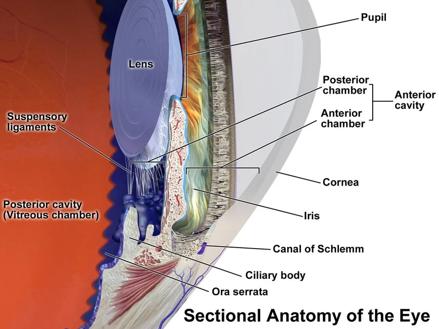
- Conjunctiva - a thin, mobile, vascular membrane covering the sclera and the inner eyelids. Its vessels blanch with topical phenylephrine and move when the conjunctiva is nudged with a cotton bud. Inflammation here produces diffuse redness, discharge and grittiness, but no true pain and no visual loss.
- Cornea - avascular, transparent and densely innervated by the ophthalmic division of the trigeminal nerve. That density of innervation is why corneal pathology hurts far out of proportion to its size, and why it causes reflex lacrimation, blepharospasm and photophobia.
- Limbus - the junction of cornea and sclera, where the deep anterior ciliary vessels sit. Inflammation inside the eye dilates these vessels and produces a ring of redness deepest at the limbus and fading outward: ciliary flush, or circumcorneal injection.
- Anterior chamber - the space between cornea and iris, filled with aqueous humour produced by the ciliary body and drained through the trabecular meshwork in the drainage angle. Inflammatory cells here, or a blocked angle, both give a painful red eye.
- Sclera and episclera - the tough white coat and the vascular layer overlying it. Episcleral inflammation is uncomfortable and harmless; scleral inflammation is deeply painful and often signals systemic vasculitis.
Causes
It is more useful to sort the causes by urgency than by anatomy, because urgency is the decision you actually have to make.
Sight-threatening: same-day ophthalmology
- Acute angle closure glaucoma - severe pain, haloes around lights, nausea and vomiting, a fixed mid-dilated oval pupil and a hard, hazy eye
- Microbial keratitis - a corneal ulcer, most often in a contact lens wearer, with a white corneal infiltrate that stains with fluorescein
- Anterior uveitis (iritis) - an aching photophobic eye with ciliary flush, a small or irregular pupil and cells in the anterior chamber
- Scleritis - boring pain that wakes the patient, deep violaceous redness that does not blanch, often with an underlying vasculitis
- Penetrating or perforating injury - a peaked pupil, shallow anterior chamber, or a positive Seidel test
- Chemical injury, particularly alkali - irrigate first and ask questions afterwards
- Endophthalmitis - profound pain and visual loss after intraocular surgery or an intravitreal injection, usually with a hypopyon
- Orbital cellulitis - proptosis, painful or restricted eye movements and systemic upset
Not usually sight-threatening
- Conjunctivitis - bacterial, viral or allergic; the commonest cause by a wide margin3
- Subconjunctival haemorrhage - a flat, painless block of blood with normal vision
- Episcleritis - sectoral redness with mild discomfort, blanching with topical phenylephrine
- Blepharitis and meibomian gland dysfunction - chronic gritty red eyes with crusted lid margins
- Dry eye disease - burning and grittiness worse through the day and in air-conditioned or windy environments
- Corneal abrasion or foreign body - painful and dramatic, but healing within days if uncomplicated5
- Stye (hordeolum) and chalazion - localised lid lesions with a quiet globe
- Pterygium and pinguecula - degenerative conjunctival lesions that intermittently inflame
Clinical features
The history usually gets you most of the way to a diagnosis before you have looked at the eye at all.
The history
- Vision - ask specifically whether vision is blurred, and whether the blurring clears on blinking. Discharge blurs vision transiently and clears with a blink; genuine visual loss does not.
- Pain - distinguish grittiness or a foreign body sensation (surface) from deep aching (uveitis, scleritis) from severe pain with nausea and vomiting (angle closure). Pain that wakes the patient at night points to scleritis.
- Photophobia - true photophobia, where light causes pain rather than dazzle, indicates corneal or intraocular inflammation. It is not a feature of simple conjunctivitis.
- Discharge - purulent suggests bacterial conjunctivitis, watery suggests viral or allergic, stringy mucus suggests allergy or dry eye
- Onset and laterality - conjunctivitis often starts in one eye and spreads to the other over a day or two; uveitis and angle closure are usually unilateral
- Contact lenses - any red eye in a contact lens wearer is microbial keratitis until proved otherwise. Ask about overnight wear, showering or swimming in lenses, and hygiene.
- Trauma and occupation - hammering, grinding, drilling or strimming raises the possibility of an intraocular foreign body, which can present with a deceptively quiet eye
- Systemic history - inflammatory back pain and HLA-B27 disease, inflammatory bowel disease, rheumatoid arthritis, sarcoidosis, recent viral illness, or a rash in the ophthalmic dermatome
Interpreting the pattern of redness
| Pattern | Description | Suggests |
|---|---|---|
| Diffuse conjunctival injection | Redness greatest in the fornices, fading towards the limbus; vessels move with a cotton bud | Conjunctivitis |
| Ciliary flush | Deep pink or violet ring densest at the limbus, fading outwards | Keratitis, anterior uveitis, acute angle closure |
| Sectoral redness | One wedge of redness, blanching with phenylephrine 2.5% | Episcleritis |
| Deep violaceous redness | Does not blanch with phenylephrine; often better appreciated in daylight than under a lamp | Scleritis8 |
| Flat uniform block of blood | Sharply demarcated, no visible vessels within it, sclera not visible through it | Subconjunctival haemorrhage |
Examination
A red eye examination in an OSCE has a fixed structure. Do it in the same order every time and it becomes hard to miss anything.
- Visual acuity, each eye separately, with distance glasses or through a pinhole. Record it as a Snellen fraction. If it improves through a pinhole the problem is refractive rather than pathological.
- Inspection in good light - lid position and swelling, discharge, the pattern of redness, and whether there is proptosis. Compare the two eyes directly.
- Pupils - size, shape, symmetry and reaction to light, including the swinging light test for a relative afferent pupillary defect. A distorted or unreactive pupil is always abnormal.
- Eye movements - painful or restricted movement suggests orbital disease rather than surface disease
- Fluorescein staining with a cobalt blue light, after a drop of topical anaesthetic if needed. Look for the linear scratch of an abrasion, the branching dendrite of herpes simplex, or the round staining infiltrate of a bacterial ulcer.
- Lid eversion if there is any suggestion of a foreign body. A subtarsal foreign body causes vertical linear corneal scratches and is easily missed.
- Fundoscopy, and where the skills and equipment exist, slit lamp examination and intraocular pressure measurement
Differential diagnosis
The following table is the core of the topic, and is worth being able to reproduce from memory.
| Diagnosis | Vision | Pain | Pupil | Redness | Other features |
|---|---|---|---|---|---|
| Bacterial conjunctivitis | Normal | Gritty | Normal | Diffuse | Purulent discharge, lids stuck together on waking |
| Viral conjunctivitis | Normal | Gritty | Normal | Diffuse | Watery discharge, follicles, tender preauricular node |
| Allergic conjunctivitis | Normal | Itch | Normal | Diffuse | Itch is the defining symptom; chemosis, atopy |
| Subconjunctival haemorrhage | Normal | None | Normal | Blood, sharply edged | Often on warfarin or a DOAC; check blood pressure |
| Episcleritis | Normal | Mild | Normal | Sectoral, blanches | Self-limiting over 1-2 weeks |
| Scleritis | May be reduced | Severe, boring, wakes the patient | Normal | Violaceous, does not blanch | Around half have a systemic vasculitis or rheumatoid arthritis |
| Anterior uveitis | Blurred | Aching, photophobia | Small, irregular | Ciliary flush | Cells and flare, posterior synechiae, hypopyon if severe |
| Microbial keratitis | Reduced | Severe | Normal or small | Ciliary flush | White corneal infiltrate staining with fluorescein; contact lenses |
| Acute angle closure | Markedly reduced, haloes | Severe, with nausea and vomiting | Mid-dilated, fixed, oval | Ciliary flush | Hazy cornea, stony hard globe, systemically unwell4 |
Investigations
Most red eyes need no investigation at all. The diagnosis is clinical, and the useful tests are performed at the bedside.
- Fluorescein staining - mandatory in any painful red eye. Also used for the Seidel test: a positive test, where fluorescein is diluted into a green stream by leaking aqueous, indicates a full-thickness corneal wound and is a surgical emergency.
- Intraocular pressure - by Goldmann applanation or a handheld tonometer. Normal is 10-21 mmHg, and angle closure typically produces pressures above 40 mmHg. Do not measure pressure if globe rupture is suspected.
- Slit lamp examination - the definitive assessment, allowing cells and flare in the anterior chamber, hypopyon, keratic precipitates and corneal infiltrates to be seen directly
- Corneal scrape for microscopy and culture - performed by ophthalmology in suspected microbial keratitis, before starting intensive topical antibiotics
- Conjunctival swabs - reserved for hyperacute, neonatal or treatment-resistant conjunctivitis. Send a charcoal swab for bacterial culture, plus a specific chlamydia and gonococcus nucleic acid amplification test where those are suspected.
- Systemic bloods - FBC, CRP, ESR, ANCA, rheumatoid factor, serum ACE, HLA-B27 and syphilis serology, guided by the pattern of recurrent uveitis or scleritis
- Imaging - orbital CT for suspected orbital cellulitis or an intraocular foreign body. Never request MRI first if a metallic foreign body is possible.
Management
Management divides into what you can do yourself, what you refer, and how urgently you refer it.
Immediate action at the bedside
Conditions you can manage in the community
- Bacterial conjunctivitis - usually self-limiting; topical chloramphenicol if severe or not settling7
- Viral conjunctivitis - supportive care, cool compresses and lubricants, with clear hygiene advice, as adenovirus remains highly contagious for up to two weeks
- Allergic conjunctivitis - allergen avoidance, a topical antihistamine, or a mast cell stabiliser such as sodium cromoglicate
- Subconjunctival haemorrhage - reassurance; check blood pressure and, if on warfarin, the INR
- Episcleritis - lubricants, with oral NSAIDs if troublesome
- Blepharitis and dry eye - lid hygiene, warm compresses and preservative-free lubricants, explaining that this is a chronic condition to be managed rather than cured
- Uncomplicated corneal abrasion - topical chloramphenicol ointment as prophylaxis, analgesia and review at 48 hours. Do not pad the eye of a contact lens wearer.
Referral thresholds
| Urgency | Presentations |
|---|---|
| Immediate (999 or straight to eye casualty) | Chemical injury, penetrating injury or suspected globe rupture, acute angle closure glaucoma, endophthalmitis, orbital cellulitis |
| Same day | Suspected microbial keratitis, any red eye in a contact lens wearer, anterior uveitis, scleritis, herpes zoster ophthalmicus, red eye with reduced acuity from any cause, hypopyon or hyphaema |
| Within a week | Recurrent episcleritis, conjunctivitis not resolving after 7-10 days of treatment, suspected chlamydial conjunctivitis, chronic red eye of unclear cause |
| Routine | Chalazion persisting after 4-6 weeks of conservative treatment, symptomatic pterygium, chronic blepharitis resistant to lid hygiene |
Special situations
The contact lens wearer
Treat every red eye in a contact lens wearer as microbial keratitis until an ophthalmologist has examined the cornea at a slit lamp. Overnight wear, swimming or showering in lenses, and topping up rather than replacing solution all increase the risk. Pseudomonas aeruginosa is the classic organism and can melt a cornea within 24 hours, while Acanthamoeba keratitis, associated with tap water exposure, causes pain far out of proportion to the signs. Advise the patient to stop lens wear immediately and to bring their lenses and case, which can be cultured.
The neonate
Conjunctivitis in the first 28 days of life is ophthalmia neonatorum and is a notifiable condition. Gonococcal infection presents within the first 48 hours with hyperacute purulent discharge and can perforate the cornea; chlamydial infection presents at 5-14 days and is associated with pneumonitis. Both need urgent paediatric and ophthalmology involvement, systemic antibiotics, and treatment of the mother and her partners.
Herpes zoster ophthalmicus
Shingles in the ophthalmic division of the trigeminal nerve threatens the eye in around half of cases. Hutchinson's sign - vesicles on the tip, side or root of the nose, reflecting involvement of the nasociliary nerve - predicts ocular involvement, although its absence does not exclude it. Start oral aciclovir 800 mg five times daily within 72 hours of rash onset and refer for slit lamp assessment.6
The child
Children are poor historians and readily squeeze the eye shut, so a red eye in a child needs patience and often a second look. Bilateral sticky eyes in an infant are usually a blocked nasolacrimal duct. A unilateral red eye with lid swelling and fever raises periorbital or orbital cellulitis, and any child who cannot open the eye, has proptosis, or has restricted movements needs immediate assessment. Uveitis in a child with juvenile idiopathic arthritis is often painless and asymptomatic, and is detected only on formal screening.
Red flags
Prognosis
For the great majority - conjunctivitis, subconjunctival haemorrhage, episcleritis and uncomplicated abrasions - the prognosis is excellent, and the eye returns to normal within days to two weeks with no lasting effect on vision.
The sight-threatening group behaves entirely differently, and the determinant of outcome is time to treatment rather than the treatment itself. Retinal ganglion cells lost during a prolonged episode of acute angle closure do not recover. A central corneal ulcer that heals leaves a scar that permanently degrades acuity, however well it is eventually treated. Alkali injury that has already caused limbal ischaemia destroys the stem cells that resurface the cornea, and the resulting conjunctivalisation is very difficult to reverse.
This asymmetry - most cases benign, a few catastrophic and time-critical - is exactly why the structured approach matters. Measuring acuity, staining with fluorescein and looking deliberately for ciliary flush takes a few minutes, and it is what converts an unstructured guess into a safe assessment.
References
- NICE Clinical Knowledge Summaries. Red eye. Available here
- Royal College of Ophthalmologists. Clinical guidelines and resources. Available here
- NICE Clinical Knowledge Summaries. Conjunctivitis - infective. Available here
- NICE Clinical Knowledge Summaries. Glaucoma. Available here
- NICE Clinical Knowledge Summaries. Corneal superficial injury. Available here
- NICE Clinical Knowledge Summaries. Shingles. Available here
- BNF. Chloramphenicol - eye preparations. Available here
- Watson PG, Hayreh SS. Scleritis and episcleritis. British Journal of Ophthalmology. 1976. Available here
This article is written for revision and education. It is not clinical guidance and must not be used to make decisions about the care of a patient. Always check current NICE guidance and local protocols.