
Radiculopathy and Peripheral Nerve Palsies: Diagnosis and Management
Key points
- Radiculopathy: dysfunction of a spinal nerve root, causing pain, sensory loss and weakness in a dermatomal/myotomal distribution, most often from disc prolapse or degenerative spinal disease.
- Peripheral nerve palsy (mononeuropathy): dysfunction of a single named peripheral nerve, most often from compression or entrapment at a specific anatomical site.
- Localising principle: a root lesion follows a dermatome/myotome that crosses multiple peripheral nerves; a peripheral nerve lesion follows that nerve's specific territory, which crosses multiple dermatomes.
- Carpal tunnel syndrome: the commonest entrapment neuropathy - median nerve compression at the wrist causing nocturnal hand pain and paraesthesiae in the radial three and a half digits.
- Common peroneal nerve palsy: foot drop with sensory loss over the dorsum of the foot, classically after fibular neck compression.
- Investigations: nerve conduction studies and electromyography localise and characterise the lesion; MRI spine images root compression.
- Management: conservative measures and physiotherapy for most; surgical decompression for red flags, severe deficit, or failure of conservative treatment.
- Cauda equina syndrome: a surgical emergency arising from multiple lumbosacral nerve roots, with bilateral leg symptoms and sphincter disturbance - not a routine radiculopathy.
Introduction
Radiculopathy and peripheral (mononeuropathy) nerve palsies are both very common presentations of focal neurological deficit, and the single most useful clinical skill in this area is distinguishing the two by pattern, since a root lesion and a peripheral nerve lesion produce genuinely different distributions of weakness and sensory loss even when they overlap anatomically.1
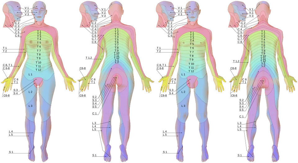
A radiculopathy affects a single nerve root as it exits the spinal canal, before it has joined the plexus and been redistributed into individual peripheral nerves - so its deficit follows a dermatome and myotome, which typically spans parts of several different peripheral nerves. A peripheral nerve palsy affects a named nerve after this redistribution, so its deficit follows that nerve's specific sensory and motor territory, which typically spans parts of several different dermatomes/roots.
Two further clues help separate the two at the bedside. The first is pain distribution: radicular pain is typically sharp and shooting, radiates in a linear band down the limb, and is aggravated by manoeuvres that raise intraspinal pressure such as coughing or straining - none of which affect a peripheral nerve lesion. The second is the presence of a specific compression point: a peripheral nerve palsy usually has a plausible mechanical cause at a known anatomical site, and tapping over it may reproduce paraesthesiae in the nerve territory (Tinel's sign).
Reflexes provide the third discriminator. A reflex arc depends on a single root level, so loss of a specific reflex - the ankle jerk in S1, the triceps in C7 - localises to that root. Most peripheral nerve palsies, by contrast, spare the common reflexes entirely, because the nerve involved does not carry the whole reflex arc.
A further principle worth holding onto is that nerve roots and peripheral nerves have a many-to-many relationship. A single root contributes fibres to several peripheral nerves, and a single peripheral nerve draws fibres from several roots. This is why an L5 radiculopathy weakens both ankle dorsiflexion (via the deep peroneal nerve) and hip abduction (via the superior gluteal nerve) - muscles supplied by entirely different peripheral nerves that happen to share a root.
Testing a muscle pair that shares a root but not a nerve, or shares a nerve but not a root, is therefore the most efficient way to localise a lesion at the bedside, and is the reasoning behind the foot drop example in the exam tip above.
Carpal tunnel syndrome is by some distance the commonest entrapment neuropathy, affecting several percent of the adult population and considerably more in specific groups such as pregnant women and those with diabetes, hypothyroidism or rheumatoid arthritis. Lumbosacral radiculopathy is likewise common, with sciatica affecting a substantial minority of adults at some point. Both are therefore conditions a junior clinician will meet regularly, and both are frequently diagnosed and managed without ever requiring imaging.
Radiculopathy
Aetiology
- Intervertebral disc prolapse - the commonest cause in younger and middle-aged adults, most often at the lumbosacral (L4/5, L5/S1) or cervical (C6/7) levels
- Degenerative spinal disease - facet joint hypertrophy and osteophyte formation narrowing the exit foramen, more common in older patients
- Spinal stenosis - can cause radiculopathy alone or combined with neurogenic claudication if multiple levels are affected
- Malignant infiltration or vertebral metastasis
- Infection - vertebral osteomyelitis or discitis compressing an adjacent root
- Diabetic radiculopathy (diabetic amyotrophy) - a painful, asymmetric proximal lower limb radiculopathy/plexopathy, sometimes with significant weight loss, that can occur even with well-controlled diabetes
Clinical features by level
| Root | Sensory distribution | Motor deficit | Reflex affected |
|---|---|---|---|
| C6 | Thumb and index finger | Elbow flexion, wrist extension | Biceps/supinator |
| C7 | Middle finger | Elbow extension, wrist flexion | Triceps |
| L4 | Medial shin/ankle | Knee extension | Knee (patellar) |
| L5 | Dorsum of the foot, big toe | Ankle dorsiflexion, big toe extension | None reliably affected (no discrete reflex) |
| S1 | Lateral foot/sole | Ankle plantarflexion | Ankle (Achilles) |
- Radicular pain - sharp, shooting pain radiating along the dermatome, often worse with movements that increase intraspinal pressure (coughing, sneezing, straining, or specific postures)
- Sensory disturbance in the corresponding dermatome
- Weakness in the corresponding myotome
- Reduced or absent reflex where a discrete reflex arc corresponds to that root
- Positive nerve root tension signs: straight leg raise (L4-S1) reproducing radicular pain, or its cervical equivalent (Spurling's test - neck extension, lateral flexion and axial compression reproducing arm symptoms)
Common peripheral nerve palsies
Peripheral (mononeuropathy) palsies most often arise from compression at a predictable anatomical site, or from trauma, and each has a recognisable signature of weakness and sensory loss that spans multiple dermatomes.
| Nerve | Common cause/site | Motor deficit | Sensory loss |
|---|---|---|---|
| Median nerve (carpal tunnel) | Compression at the wrist (carpal tunnel syndrome) - pregnancy, hypothyroidism, rheumatoid arthritis, acromegaly, repetitive strain all predispose | Thumb abduction/opposition (thenar wasting in longstanding cases) | Radial three and a half digits (palmar aspect) |
| Ulnar nerve | Compression at the elbow (cubital tunnel) - prolonged leaning on the elbow, or fracture/deformity | Finger abduction/adduction (interossei), claw hand in longstanding cases | Little finger and medial half of ring finger |
| Radial nerve | Compression against the humerus ('Saturday night palsy' from prolonged arm compression, or humeral fracture) | Wrist and finger extension - wrist drop | Dorsum of the hand (first dorsal web space) |
| Common peroneal nerve | Compression at the fibular neck (crossing legs, prolonged kneeling, tight casts) | Ankle dorsiflexion and eversion - foot drop, with a high-stepping gait | Dorsum of the foot and lateral shin |
| Femoral nerve | Compression (pelvic mass, retroperitoneal haemorrhage - consider in anticoagulated patients), hip surgery | Knee extension, hip flexion | Anterior thigh and medial shin |
| Lateral cutaneous nerve of the thigh (meralgia paraesthetica) | Compression under the inguinal ligament - obesity, tight clothing, pregnancy | None (purely sensory) | Anterolateral thigh |
Clinical examination
- Precise mapping of sensory loss - dermatomal (crossing multiple peripheral nerve territories) versus a single peripheral nerve's specific territory
- Precise mapping of motor deficit - myotomal versus a single nerve's specific muscle group
- Reflexes - a specific reflex loss supports a root lesion at that level
- Provocation tests - straight leg raise, Spurling's test, Tinel's and Phalen's tests for carpal tunnel syndrome, Tinel's sign at the elbow for ulnar neuropathy
- Look for the precipitant - a specific compression point, recent trauma, prolonged awkward posture, or systemic disease predisposing to entrapment (diabetes, hypothyroidism, rheumatoid arthritis, pregnancy, acromegaly)
- Assess for signs pointing to a more serious cause - bilateral symptoms, sphincter disturbance, systemic symptoms (weight loss, fever), or rapidly progressive weakness
Differential diagnosis
- Cauda equina syndrome - multiple lumbosacral roots affected bilaterally, with saddle anaesthesia and sphincter disturbance; a surgical emergency, not a routine radiculopathy (see red flags)
- Spinal cord compression - upper motor neurone signs below the level of compression, a sensory level, and sphincter involvement, distinguishing it from a single root or peripheral nerve lesion
- Plexopathy (brachial or lumbosacral) - affects multiple roots/nerves in a pattern that does not fit a single root or single peripheral nerve, from trauma, malignant infiltration, or radiation3
- Peripheral polyneuropathy - symmetric, length-dependent, and typically bilateral, unlike the usually unilateral, focal pattern of a single radiculopathy or mononeuropathy
- Mononeuritis multiplex - sequential involvement of multiple individual nerves, a red flag for vasculitis (covered in Peripheral Neuropathy)
Investigations
- Clinical diagnosis is usually sufficient for classic, mild-to-moderate presentations of both radiculopathy and common entrapment neuropathies, and imaging/neurophysiology is reserved for atypical features, red flags, or planning for intervention
- MRI spine - for radiculopathy with red flags, significant or progressive weakness, or failure to improve with conservative management, to identify disc prolapse, foraminal stenosis, or a more sinister cause (tumour, infection)
- Nerve conduction studies and electromyography - localise and characterise a peripheral nerve palsy, confirm the diagnosis of, for example, carpal tunnel syndrome, and distinguish it from a root or plexus lesion when the clinical picture is ambiguous
- Ultrasound - increasingly used to visualise nerve entrapment directly, particularly for carpal tunnel syndrome
- Screen for predisposing systemic causes - HbA1c, TFTs, rheumatoid factor, particularly for carpal tunnel syndrome without an obvious mechanical cause
Management
Radiculopathy
- Analgesia - paracetamol and NSAIDs first-line; neuropathic agents (amitriptyline, gabapentin) for persistent radicular pain
- Physiotherapy and continued activity as tolerated - prolonged bed rest is not recommended and worsens outcomes
- Epidural corticosteroid injection - can provide short-to-medium-term relief in selected patients with persistent radicular pain
- Surgical decompression (discectomy or foraminotomy) - considered for significant or progressive motor deficit, or pain that fails to settle with adequate conservative management (generally trialled for at least 6-12 weeks unless red flags are present)
Peripheral nerve palsies
- Remove or modify the precipitant - wrist splinting (particularly at night) for carpal tunnel syndrome, avoiding leaning on the elbow for ulnar neuropathy, padding/positioning to avoid fibular neck compression
- Corticosteroid injection - for carpal tunnel syndrome not settling with splinting alone
- Treat the underlying predisposing condition - hypothyroidism, diabetes, rheumatoid arthritis
- Surgical decompression - carpal tunnel release for persistent or severe carpal tunnel syndrome (especially with thenar wasting or persistent sensory loss)2, cubital tunnel decompression for refractory ulnar neuropathy
- Physiotherapy and splinting to prevent contracture and support function during recovery, particularly for foot drop (ankle-foot orthosis) or wrist drop
Complications
- Persistent pain and sensory disturbance if decompression is delayed
- Permanent motor deficit or muscle wasting from prolonged, severe compression
- Contractures (for example, an untreated foot drop leading to fixed equinus deformity)
- Falls and injury from foot drop or other motor deficits affecting gait
- Missed cauda equina syndrome or spinal cord compression if red flags are not actively sought
Red flags
The great majority of radiculopathies and entrapment neuropathies are benign and self-limiting, so the purpose of the features below is to identify the small minority that are not - particularly cauda equina syndrome, where delay causes irreversible harm.
Prognosis
Most radiculopathies from disc prolapse improve substantially with conservative management within weeks to a few months, as the extruded disc material resorbs and inflammation settles. Common entrapment neuropathies such as carpal tunnel syndrome often respond well to splinting or, if needed, decompression, particularly when treated before significant wasting develops - established thenar or other muscle wasting recovers more slowly and sometimes incompletely. Diabetic radiculopathy/amyotrophy, though initially alarming, usually improves gradually over many months even without specific treatment, though recovery can be prolonged and sometimes incomplete.
A final practical point is that the natural history of most radiculopathies is favourable, and this should be communicated clearly. Explaining that the majority improve substantially within six to twelve weeks, that imaging findings correlate poorly with symptoms and often show incidental degenerative change in asymptomatic people, and that remaining active is beneficial rather than harmful, does more to aid recovery than any specific intervention. Unnecessary early imaging frequently leads to over-treatment of findings that were never the cause of the pain.
References
- NICE NG59. Low back pain and sciatica in over 16s: assessment and management. 2016, updated 2020. Available here
- NICE CKS. Carpal tunnel syndrome. Available here
- Wilbourn AJ. Plexopathies. Neurologic Clinics. 2007. Available here
This article is written for revision and education. It is not clinical guidance and must not be used to make decisions about the care of a patient. Always check current NICE guidance and local protocols.