
Infectious Mononucleosis
Key points
- Infectious mononucleosis: usually caused by primary infection with Epstein-Barr virus (EBV), spread in saliva and classically affecting teenagers and young adults.
- Classic triad: fever, exudative pharyngotonsillitis, and lymphadenopathy - characteristically involving the posterior cervical chain.
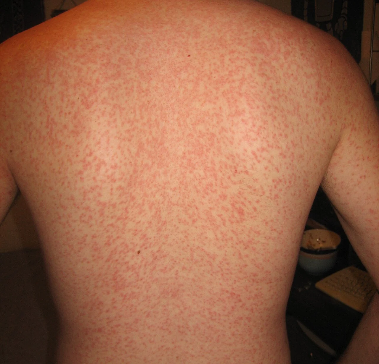
- The amoxicillin rash: giving amoxicillin or ampicillin for the sore throat provokes a widespread, itchy maculopapular rash in a large majority of patients - it is not a marker of true penicillin allergy.
- Diagnosis: blood film showing lymphocytosis with atypical lymphocytes, plus a positive heterophile antibody (Monospot/Paul-Bunnell) test; EBV-specific serology if the heterophile test is negative or the patient is young.
- Management: supportive - rest, fluids, paracetamol/NSAIDs; antibiotics have no role against the virus itself.
- Splenic rupture: the reason for advising against contact sport and heavy lifting for at least 3-4 weeks - splenomegaly is common and the enlarged spleen is vulnerable to even minor trauma.
- Prognosis: excellent in the great majority, though fatigue can persist for weeks to months after the acute illness has resolved.
Introduction
Infectious mononucleosis ("glandular fever") is most often caused by primary infection with the Epstein-Barr virus (EBV), a member of the herpesvirus family, spread through saliva - hence its colloquial name, the "kissing disease". Most primary EBV infection in early childhood is mild or asymptomatic; the classic florid illness is seen mainly when primary infection occurs in adolescence or young adulthood.1
A clinically identical picture can be caused by cytomegalovirus (CMV), and less commonly by toxoplasmosis, acute HIV seroconversion, or adenovirus - together sometimes grouped as "heterophile-negative" mononucleosis, since the specific antibody test used for EBV is negative in these cases. Like all herpesviruses, EBV establishes lifelong latency (in B lymphocytes) after primary infection, with the potential to reactivate under significant immunosuppression.
Pathophysiology
EBV infects oropharyngeal epithelial cells and then B lymphocytes, driving polyclonal B-cell activation. The dominant host response is a vigorous CD8+ cytotoxic T-cell expansion against EBV-infected B cells - it is this reactive T-cell population, not the infected B cells themselves, that produces the atypical lymphocytes seen on the blood film, and that accounts for much of the lymphadenopathy and systemic illness.
Clinical features
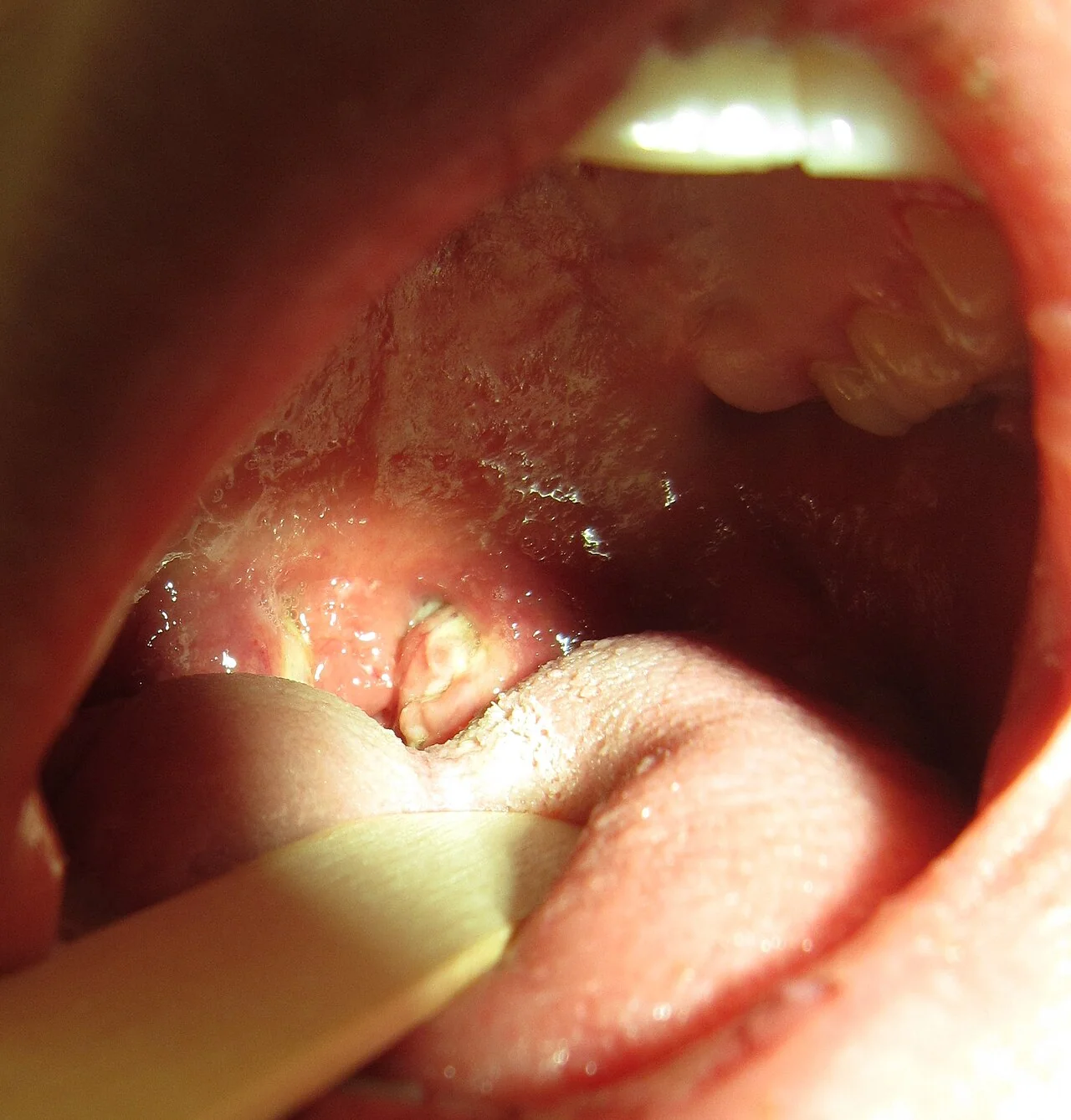
The classic triad is fever, severe exudative pharyngotonsillitis, and lymphadenopathy, developing over several days and typically lasting 2-4 weeks.
- Fever, often high and prolonged compared with a routine viral sore throat
- Exudative pharyngotonsillitis - can be dramatic, with thick white-grey exudate, and is easily mistaken for bacterial tonsillitis or, in severe cases, quinsy
- Lymphadenopathy - generalised, but posterior cervical chain involvement is a useful discriminator from bacterial tonsillitis, which more typically produces anterior cervical nodes
- Marked fatigue and malaise, often out of proportion to the other findings, and sometimes the most prominent complaint
- Splenomegaly - present in around half of patients
- Hepatomegaly and mild hepatitis - deranged transaminases are common, usually mild and self-limiting
- Palatal petechiae
- Periorbital or eyelid oedema (Hoagland's sign) - a less common but recognised early feature
Clinical examination
- Inspect the oropharynx for tonsillar enlargement and exudate, and assess for any airway compromise if swelling is severe
- Palpate all lymph node groups, noting posterior cervical involvement specifically
- Palpate for splenomegaly and hepatomegaly, gently, given the risk of splenic injury
- Check for palatal petechiae and periorbital oedema
- Look for jaundice, though this is uncommon
Differential diagnosis
- Streptococcal tonsillitis - use the Centor or FeverPAIN criteria; anterior rather than posterior cervical lymphadenopathy and a shorter course favour this over mononucleosis
- CMV mononucleosis syndrome - clinically similar but heterophile-antibody negative
- Acute HIV seroconversion illness - fever, pharyngitis, lymphadenopathy and a rash overlap closely; consider HIV testing where risk factors are present or the heterophile test is negative
- Toxoplasmosis - a similar glandular fever-like picture, particularly relevant with cat or undercooked meat exposure
- Diphtheria - rare in the UK, but a classic historical differential given the pseudomembranous exudate and systemic illness
- Viral hepatitis - if hepatomegaly and deranged LFTs dominate the picture
Investigations
- FBC and blood film - a lymphocytosis, with atypical (reactive) lymphocytes exceeding around 10% of the total white cell count, is characteristic
- Heterophile antibody test (Monospot/Paul-Bunnell test) - the standard rapid test; can be falsely negative in the first week of illness and is less reliable in children under about 12, so a negative result early on should be repeated after 1-2 weeks if suspicion remains high
- EBV-specific serology - useful when the heterophile test is negative or unreliable: VCA IgM indicates acute infection, VCA IgG indicates current or past infection, and EBNA IgG, which only appears several weeks into convalescence, helps confirm that an infection is not recent
- LFTs - mildly deranged transaminases are common and do not usually need specific action
- Throat swab - if there is genuine diagnostic uncertainty with bacterial tonsillitis, though clinical judgement (and avoiding amoxicillin/ampicillin until the picture is clearer) usually suffices
Management
Management is supportive, since there is no specific antiviral treatment in routine use for uncomplicated disease.
- Rest, adequate fluid intake, and paracetamol or NSAIDs for fever and pain
- Avoid amoxicillin or ampicillin for the sore throat, both because they will not help a viral illness and because of the well-known rash reaction described above
- Avoid contact sports, heavy lifting and other significant physical exertion for at least 3-4 weeks, and until any splenomegaly has been confirmed to have resolved, because of the risk of splenic rupture
- Corticosteroids are reserved for specific complications - for example, impending airway obstruction from severe tonsillar swelling, or significant autoimmune haemolytic anaemia or thrombocytopenia - rather than used routinely
Complications
- Splenic rupture - rare, but the most feared complication, presenting with left upper quadrant or referred left shoulder tip pain (Kehr's sign) and haemodynamic compromise; a surgical emergency
- Upper airway obstruction from severe tonsillar hypertrophy, occasionally needing corticosteroids or ENT intervention
- Hepatitis, usually mild and self-limiting; fulminant hepatic failure is very rare
- Autoimmune haemolytic anaemia, thrombocytopenia and, rarely, neutropenia
- Neurological complications - Guillain-Barré syndrome, encephalitis and meningitis, all uncommon
- Chronic fatigue persisting for weeks to months after the acute illness has otherwise resolved
- Long-term oncogenic potential - EBV is associated with Burkitt lymphoma, Hodgkin lymphoma, nasopharyngeal carcinoma, and post-transplant lymphoproliferative disease in immunosuppressed patients, reflecting its lifelong latency in B cells
Red flags
Prognosis
The great majority of patients make a full recovery, with acute symptoms settling over 2-4 weeks. Fatigue is often the slowest symptom to resolve and can persist for weeks to several months, which is worth explaining early so that a prolonged recovery is not mistaken for a new or ongoing problem. Serious complications are uncommon but are the reason behind the standard safety-netting advice on avoiding contact sport and seeking urgent review for severe abdominal pain.
References
- NICE Clinical Knowledge Summaries. Glandular fever (infectious mononucleosis). Available here
- Dunmire SK, Hogquist KA, Balfour HH. Infectious Mononucleosis. Current Topics in Microbiology and Immunology. 2015. Available here
- Luzuriaga K, Sullivan JL. Infectious mononucleosis. New England Journal of Medicine. 2010. Available here
- British Association for Sexual Health and HIV / NICE Clinical Knowledge Summaries. Sore throat - acute. Available here
This article is written for revision and education. It is not clinical guidance and must not be used to make decisions about the care of a patient. Always check current NICE guidance and local protocols.