
Perforated Tympanic Membrane: Assessment and Management
Key points
- Perforated tympanic membrane: a defect in the eardrum, most often caused by infection (acute otitis media) or trauma.
- Aetiology: infective (pressure rupture during acute otitis media) or traumatic (direct injury, barotrauma, blast injury, iatrogenic).
- Presentation: sudden otalgia (often relieved once the drum ruptures), conductive hearing loss, and bleeding or discharge from the ear.
- Examination: otoscopy showing the perforation - its size, position, and whether the edges are clean or ragged - and formal hearing testing.
- Management: conservative: keep the ear dry, avoid instrumentation, and review at 6-8 weeks, since most perforations heal spontaneously.
- Persistent perforation: one not healed by around 3 months is referred to ENT for consideration of myringoplasty.
- Complications: recurrent infection, persistent conductive hearing loss, and rarely implantation cholesteatoma.
- Red flags: signs of temporal bone fracture (CSF otorrhoea, haemotympanum, Battle's sign) or vertigo/facial weakness need urgent imaging.
Introduction
A perforated tympanic membrane is a defect in the eardrum that breaches the barrier between the external canal and the middle ear. It is a common finding in both primary care and emergency medicine, arising either as a complication of infection or from trauma to the ear.1
The great majority heal spontaneously with simple conservative measures, so the key clinical tasks are confirming the diagnosis, excluding a more serious underlying cause such as temporal bone fracture, and knowing when a perforation has failed to heal and needs surgical referral.
Aetiology and pathophysiology
Infective
The most common cause is acute otitis media, where a build-up of pus under pressure in the middle ear ruptures the drum, most often at its weakest, most vascular point (the pars tensa). This typically brings sudden relief of the preceding severe ear pain, followed by blood-stained or purulent discharge.1
Traumatic
- Direct trauma: cotton buds, hairgrips, or other objects pushed into the canal
- Barotrauma: a sudden pressure differential across the drum, from diving, flying (especially with concurrent eustachian tube dysfunction), or a direct slap to the ear
- Blast injury: explosions or very loud noise close to the ear
- Iatrogenic: ear syringing, grommet insertion, or as a complication of ear surgery
- Head injury: a temporal bone fracture can tear the drum and is associated with more serious injury
Traumatic perforations are usually more irregular and can involve tearing of the drum edges, in contrast to the more central, rounded defect typical of an infective rupture.
Risk factors
- Acute otitis media, particularly if untreated or severe
- Use of cotton buds or other objects to clean the ear
- Diving, flying with an active upper respiratory tract infection, or other barotrauma exposure
- Occupational or recreational noise exposure to blast or explosion
- Previous grommet insertion
- Head trauma
Clinical features
The presentation depends on the cause. In an infective perforation, there is often a preceding history of severe ear pain that suddenly eases as the drum ruptures, followed by discharge that may be blood-stained then mucopurulent.1 In traumatic perforation, the patient reports a sudden sharp pain at the moment of injury, immediately followed by hearing loss, and sometimes tinnitus or mild dizziness.
Conductive hearing loss is expected with any perforation, roughly proportional to its size. Bleeding from the canal is common immediately after traumatic injury. Ask specifically about the mechanism of injury (syringing, slap, explosion, diving, head trauma), since this affects both the differential and the need for further imaging.
Examination
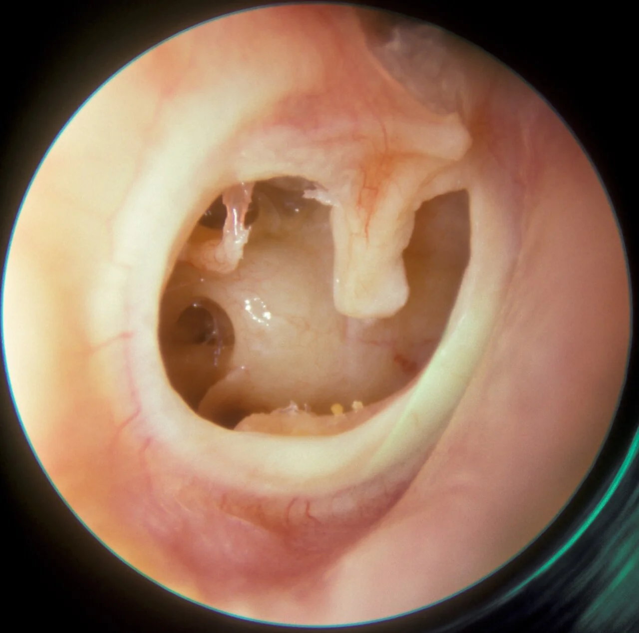
Otoscopy confirms the diagnosis, showing a visible defect in the tympanic membrane. Describe its size (small, moderate, large or subtotal), position (central/pars tensa versus marginal, which raises concern for cholesteatoma) and the state of the edges (clean in a healing perforation, ragged or everted in a fresh traumatic one).1
Tuning fork tests (Rinne and Weber) confirm a conductive hearing loss on the affected side: Rinne negative (bone conduction louder than air conduction) on the perforated ear, with Weber lateralising to that side. Formal audiometry quantifies the loss and is useful for monitoring recovery.
After significant head trauma, examine for signs of a temporal bone fracture: haemotympanum, CSF otorrhoea (a halo sign on filter paper, or testing for beta-2 transferrin), bruising over the mastoid (Battle's sign), and facial nerve function, since fractures can injure the nerve as it runs through the temporal bone.
Differential diagnosis
- Acute otitis media without perforation: intact but bulging drum
- Chronic suppurative otitis media / cholesteatoma: a marginal or attic perforation with chronic, foul discharge rather than an acute event
- Grommet in situ: a small, round, deliberately placed opening that should not be mistaken for pathology
- Otitis externa: canal inflammation without a drum defect
Investigations
Most perforations are diagnosed clinically and need no further investigation beyond otoscopy and a hearing assessment.1 Audiometry documents the degree of conductive loss and provides a baseline to track healing.
CT temporal bones is indicated after significant head trauma, or when there are features suggesting a temporal bone fracture or ossicular disruption, to define the injury and plan any surgical intervention. It is not required for a straightforward infective or minor traumatic perforation.
Management
The majority of perforations, whatever the cause, heal spontaneously within a few weeks as the squamous epithelium regenerates across the defect. Initial management is therefore conservative: keep the ear dry (no swimming, and cover the ear with a vaselined cotton wool ball or ear plug while showering), avoid inserting anything into the canal, and avoid forceful nose blowing, which can push air and secretions through the eustachian tube into the middle ear.1
Antibiotics are not routinely needed for an uncomplicated perforation. If there is active discharge suggesting infection, a topical antibiotic is used - ciprofloxacin drops are preferred over aminoglycosides in this setting because the middle ear is directly exposed through the perforation, removing the barrier that normally limits ototoxic absorption.2 Oral antibiotics are reserved for associated acute otitis media with systemic symptoms.
Review is arranged at 6-8 weeks to check healing. A perforation still present at around 3 months is considered persistent and should be referred to ENT, where myringoplasty (surgical repair, usually with a graft of the patient's own fascia or cartilage) can be considered, particularly if hearing loss is significant or infections are recurrent.
Complications
- Recurrent otitis media: water or debris entering the middle ear through the open perforation
- Persistent conductive hearing loss: especially with larger or non-healing perforations
- Chronic suppurative otitis media: ongoing discharge through a perforation that fails to heal
- Implantation (secondary acquired) cholesteatoma: squamous epithelium growing into the middle ear through the perforation edge, a rare but important long-term complication
- Tinnitus: can persist even after the perforation has healed
Red flags
Prognosis
Around 90% of perforations, whether infective or traumatic, heal spontaneously within a few weeks to a couple of months without any intervention.1 Larger perforations, and those affecting a previously scarred or already-thin drum, are less likely to heal on their own and more often need surgical repair.
Myringoplasty has a high success rate, with most series reporting closure in well over 90% of appropriately selected cases, and hearing typically improves once the drum is closed, although some residual loss can remain if there has been associated ossicular damage.
References
- NICE Clinical Knowledge Summaries (CKS). Perforated eardrum. 2023. Available here
- BNF. Ear conditions - infection (ototoxicity considerations). Available here
- Michael Hawke MD, CC BY-SA 4.0, via Wikimedia Commons. Available here
This article is written for revision and education. It is not clinical guidance and must not be used to make decisions about the care of a patient. Always check current NICE guidance and local protocols.