
Acute Mastoiditis: Diagnosis and Management
Key points
- Acute mastoiditis: spread of infection from the middle ear into the mastoid air cells, the most common serious complication of acute otitis media.
- Aetiology: the same organisms as acute otitis media - Streptococcus pneumoniae, Streptococcus pyogenes, Staphylococcus aureus and Haemophilus influenzae.
- Presentation: fever and worsening ear pain following (or during) an episode of otitis media, with post-auricular swelling, erythema and tenderness.
- Key sign: a protruding, inferolaterally displaced pinna from a subperiosteal abscess is the classic examination finding.
- Investigations: clinical diagnosis supported by bloods; CT temporal bones with contrast if diagnostic doubt or a complication is suspected.
- Management: admission for IV antibiotics and ENT input; myringotomy to drain the middle ear, with mastoidectomy reserved for abscess or non-response.
- Complications: subperiosteal abscess, facial nerve palsy, labyrinthitis, and intracranial spread (meningitis, sinus thrombosis, abscess).
- Prognosis: excellent with prompt treatment; most children recover fully with IV antibiotics without needing mastoid surgery.
Introduction
Acute mastoiditis is infection and inflammation of the mastoid air cell system, almost always arising as a complication of inadequately treated or unusually aggressive acute otitis media (AOM).1 It is the most common serious complication of AOM and is predominantly a disease of young children, in whom the mastoid is still pneumatising.
Although uncommon in the era of antibiotics, it remains an important diagnosis because untreated infection can progress rapidly to abscess formation and, from there, to intracranial spread. Any child with a protruding, tender, erythematous ear and fever needs urgent assessment.
Aetiology and pathophysiology
The mastoid air cells communicate with the middle ear via the aditus ad antrum, so any middle ear infection has direct access to the mastoid. In most children this communication allows free drainage and ventilation, but during AOM the mucosa lining the mastoid becomes inflamed in parallel with the middle ear.
If the aditus becomes obstructed by inflammatory swelling, secretions and pus accumulate within the air cells under pressure. This causes periostitis, then osteitis with breakdown of the bony septa between air cells (coalescent mastoiditis), and pus can subsequently track through the thin lateral mastoid cortex to form a subperiosteal abscess beneath the skin over the mastoid.1,2
The causative organisms mirror those of acute otitis media: Streptococcus pneumoniae, Streptococcus pyogenes, Staphylococcus aureus and Haemophilus influenzae are most common, with Pseudomonas aeruginosa more typical of chronic or cholesteatoma-associated disease.
Masked mastoiditis
A distinct pattern, masked mastoiditis, can occur when a course of oral antibiotics for AOM is enough to partially suppress the infection without eradicating it. The child's ear symptoms and fever settle only partially, and low-grade mastoid inflammation smoulders behind a relatively unremarkable-looking tympanic membrane, making the diagnosis easy to miss unless it is specifically considered in a child with a prolonged, atypical illness after treated otitis media.
Risk factors
- Young age, typically under 2 years, when the mastoid air cell system is still developing
- Current or recent acute otitis media, especially if inadequately treated or recurrent
- Delayed presentation or delayed initiation of antibiotics
- Immunocompromise
- Underlying cholesteatoma, which predisposes to chronic mastoiditis
- Craniofacial abnormalities affecting eustachian tube function
Clinical features
Mastoiditis typically develops in a child with a current or recent episode of AOM who fails to improve, or worsens after initial improvement. Fever, increasing otalgia, and systemic upset (irritability, lethargy, poor feeding) are common, and the child is usually visibly unwell.1
The defining local features are post-auricular swelling, erythema and tenderness. As a subperiosteal abscess develops, the pinna is characteristically pushed inferiorly and laterally ("proptosis" of the ear), and the post-auricular skin becomes fluctuant. Otorrhoea may or may not be present, depending on whether the tympanic membrane has perforated.
Examination
Inspect for post-auricular erythema, swelling and pinna displacement before touching the ear, then gently palpate for tenderness and fluctuance over the mastoid process. Otoscopy usually shows a bulging or perforated tympanic membrane consistent with AOM, though the two conditions do not always correlate exactly in severity.1
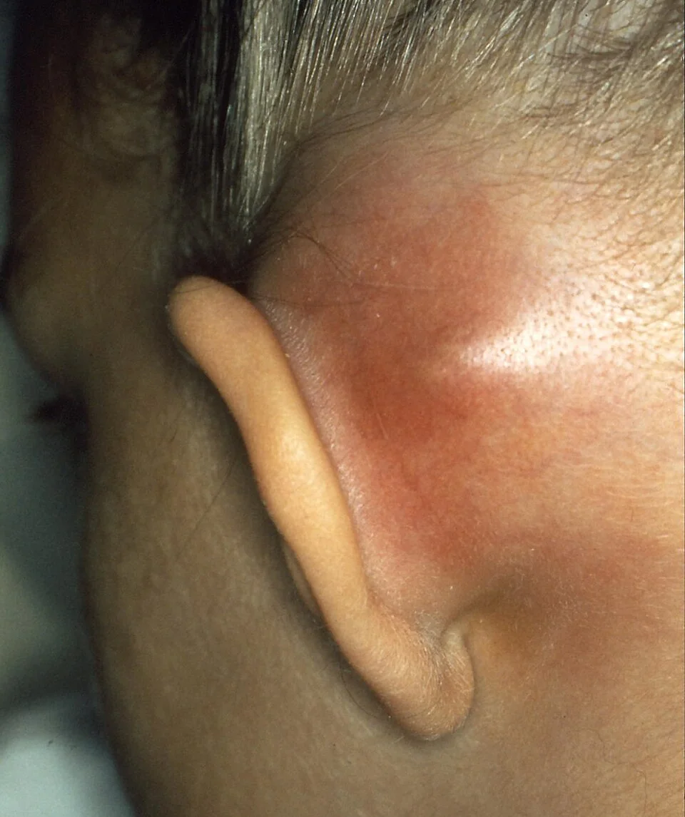
Examine facial nerve function and check for nystagmus or unsteadiness, which would suggest facial nerve involvement or labyrinthine spread respectively. A full neurological assessment, including for meningism, is essential in any child who looks more unwell than the local findings alone would suggest.
Differential diagnosis
- Post-auricular lymphadenitis: tender swelling but the pinna is not displaced and there is usually no preceding severe otitis media
- Otitis externa with peri-auricular cellulitis: canal tenderness on tragal pressure is more prominent, and the pinna is not typically pushed forward
- Infected pre-auricular sinus: swelling anterior to the ear rather than posterior, often with a visible sinus opening
- Simple acute otitis media without mastoid involvement: no post-auricular swelling, erythema or pinna displacement
Investigations
Mastoiditis is primarily a clinical diagnosis based on the history and examination findings. FBC and CRP typically show a raised white cell count and inflammatory markers, and blood cultures should be taken before starting antibiotics, particularly in a systemically unwell child.1
CT temporal bones with contrast is the investigation of choice when the diagnosis is uncertain, when a subperiosteal or intracranial abscess is suspected, or when the child is not responding to treatment as expected. It demonstrates coalescence of the mastoid air cells (loss of the normal bony septa) and can identify abscess formation or intracranial extension.2 MRI with venography is added if sigmoid sinus thrombosis or another intracranial complication is suspected.
| Feature | Otitis media alone | Acute mastoiditis | Coalescent mastoiditis |
|---|---|---|---|
| Post-auricular findings | None | Tenderness, erythema, swelling | Fluctuant abscess, pinna displaced |
| Systemic upset | Mild-moderate | Moderate-severe | Severe |
| CT mastoid air cells | Not indicated | Opacified but septa preserved | Bony septa destroyed, confluent cavity |
| First-line management | Analgesia +/- oral antibiotics | Admission, IV antibiotics, myringotomy | IV antibiotics plus surgical drainage |
Management
All children with suspected acute mastoiditis should be admitted for joint ENT and paediatric care. Initial management is with intravenous broad-spectrum antibiotics (e.g. IV co-amoxiclav or a third-generation cephalosporin, per local protocol) covering the typical AOM organisms, alongside analgesia and supportive care.1
Myringotomy (a small incision in the tympanic membrane) is usually performed to drain the middle ear, relieve pressure, and obtain a sample for culture to guide antibiotic choice; a grommet may be inserted at the same time to maintain drainage.
Many cases resolve with IV antibiotics and myringotomy alone. Surgical mastoidectomy is reserved for children with a subperiosteal abscess requiring drainage, those who fail to improve on IV antibiotics within 24-48 hours, or those with evidence of an intracranial complication.1,2
Complications
Complications reflect the routes by which infection can spread beyond the mastoid, and are the reason for urgent treatment:1,2
- Subperiosteal abscess: the most common complication, producing the classic protruding ear
- Bezold's abscess: pus tracking through the mastoid tip into the neck along the sternocleidomastoid, presenting as neck swelling
- Facial nerve palsy: from involvement of the nerve as it passes close to the mastoid
- Labyrinthitis: vertigo and sensorineural hearing loss from spread to the inner ear
- Petrous apicitis (Gradenigo's syndrome): the classic triad of otorrhoea, deep facial/retro-orbital pain (trigeminal nerve) and a lateral rectus (VI nerve) palsy causing diplopia
- Intracranial spread: meningitis, extradural or subdural abscess, brain abscess and lateral (sigmoid) sinus thrombosis are rare but life-threatening
Red flags
Prognosis
With prompt recognition and treatment, the prognosis for acute mastoiditis is excellent, and the majority of children make a full recovery without needing surgery beyond myringotomy. Serious complications are rare in developed healthcare settings but can be severe when they occur, which is why any child with the classic features should be assessed and treated without delay rather than managed expectantly as simple otitis media.1
References
- NICE Clinical Knowledge Summaries (CKS). Otitis media - acute (complications). 2023. Available here
- Laulajainen-Hongisto A, Aarnisalo AA, Jero J. Update on acute otitis media and mastoiditis in children. Advances in Oto-Rhino-Laryngology. 2020. Available here
- B. Welleschik, CC BY-SA 3.0, via Wikimedia Commons. Available here
This article is written for revision and education. It is not clinical guidance and must not be used to make decisions about the care of a patient. Always check current NICE guidance and local protocols.