
Cholesteatoma: Diagnosis and Management
Key points
- Cholesteatoma: an abnormal collection of keratinising squamous epithelium within the middle ear or mastoid, locally destructive but not a neoplasm.
- Aetiology: usually acquired, from a chronic attic retraction pocket driven by long-standing eustachian tube dysfunction; rarely congenital.
- Presentation: chronic, painless, foul-smelling discharge unresponsive to standard treatment, with progressive conductive hearing loss.
- Examination: an attic retraction pocket or perforation filled with white keratin debris, best seen with microotoscopy.
- Investigations: pure tone audiometry for hearing loss, and CT temporal bones to define disease extent before surgery.
- Management: surgery (mastoidectomy) is the only definitive treatment; there is no effective medical cure.
- Complications: erosion into the ossicles, labyrinth, facial nerve canal or dura, causing hearing loss, vertigo, facial palsy or intracranial infection.
- Prognosis: good with surgical clearance, but recurrence is common and lifelong follow-up is often needed.
Introduction
Cholesteatoma is an abnormal growth of keratinising squamous epithelium within the middle ear cleft or mastoid. Despite the name, it is neither a tumour of cholesterol nor a malignancy - it behaves like locally invasive skin in the wrong place, shedding keratin debris that becomes chronically infected and progressively erodes the surrounding bone.1
It matters clinically because, left untreated, it can destroy the ossicular chain, erode into the inner ear or facial nerve canal, and breach the skull base to cause intracranial infection. Unlike most other causes of chronic ear discharge, the only effective treatment is surgical, which makes early recognition and referral important.
Aetiology and pathophysiology
Acquired cholesteatoma (primary)
The great majority of cases are acquired. Chronic eustachian tube dysfunction causes persistently negative middle ear pressure, which draws the weakest part of the tympanic membrane - the pars flaccida, above the lateral process of the malleus - inward to form a retraction pocket. Once formed, the pocket accumulates desquamated keratin that it can no longer shed normally, and this debris becomes infected, driving further inflammation, osteoclast activation and bone erosion.1,2
Acquired cholesteatoma (secondary)
Squamous epithelium can also be directly implanted into the middle ear through a pre-existing marginal tympanic membrane perforation, or introduced iatrogenically during previous ear surgery, forming a secondary acquired cholesteatoma.
Congenital cholesteatoma
A rarer form arises from an embryonic epithelial rest trapped medial to an intact, normal-looking tympanic membrane, classically seen as a white mass behind an intact anterosuperior drum in a young child with no history of ear infection or surgery.2
Whatever the origin, the pathology is the same: keratin debris accumulates, becomes chronically infected, and the combination of pressure effects and inflammatory bone resorption erodes adjacent structures - the ossicles first, then the mastoid, the bony labyrinth, the facial nerve canal, and ultimately the tegmen (the bony plate separating the middle ear from the middle cranial fossa).
Risk factors
- Chronic or recurrent otitis media and eustachian tube dysfunction
- Cleft palate and other craniofacial abnormalities predisposing to eustachian tube dysfunction
- Previous tympanic membrane perforation or ear surgery (secondary acquired disease)
- Childhood onset, though it can present at any age
- Family history in some congenital cases
Clinical features
The classic presentation is chronic, painless, foul-smelling otorrhoea that persists or recurs despite courses of topical or oral antibiotics - a history of discharge "that never quite goes away" should always raise suspicion. Discharge may be scanty and only intermittently noticed.1
Progressive conductive hearing loss develops as keratin debris and erosion disrupt the ossicular chain, and this is often slowly progressive enough that patients and families do not seek help early. Unlike acute otitis media, pain is not a typical early feature, and its presence often signals a complication.
Later or complicated presentations include vertigo (from erosion into the lateral semicircular canal, causing a labyrinthine fistula), facial weakness (from erosion of the facial nerve canal), or signs of intracranial infection if disease has breached the skull base.2
Examination
Otoscopy classically shows an attic retraction pocket - a defect in the pars flaccida, superior to the malleus handle - filled with white, cheesy keratin debris, sometimes with surrounding granulation tissue or a crust. A posterosuperior marginal perforation with similar debris is also seen in secondary acquired disease.1
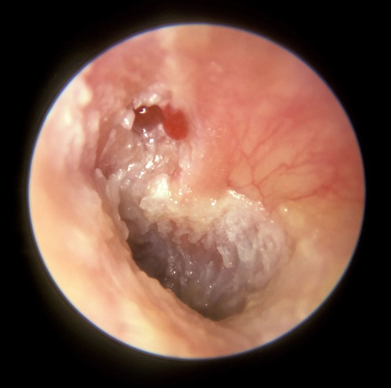
Microotoscopy or endoscopy, and aural microsuction of debris, are usually needed to fully assess the extent of a retraction pocket, since ordinary otoscopy can underestimate disease hidden deep within it. A fistula test (applying pressure to the tragus or using pneumatic otoscopy while watching for nystagmus or dizziness) is performed if a labyrinthine fistula is suspected, and facial nerve function must always be documented.
Differential diagnosis
- Chronic suppurative otitis media (without cholesteatoma): a central, non-marginal perforation with mucoid discharge
- Otitis externa: canal-based infection with tragal tenderness, not attic debris
- Aural polyp: granulation tissue protruding through a perforation, which can itself be a marker of underlying cholesteatoma and needs biopsy or exploration
- Glomus tympanicum: a vascular middle ear mass seen as a red-blue pulsatile swelling behind an intact drum
- Keratosis obturans: accumulated keratin plugging the external canal, causing canal widening rather than middle ear erosion
Investigations
Pure tone audiometry quantifies the conductive hearing loss and provides a baseline before surgery. A significant air-bone gap is typical, reflecting ossicular chain involvement.
CT of the temporal bones is the key imaging investigation for surgical planning, showing soft tissue filling the middle ear and mastoid, and demonstrating bony erosion of the ossicles, the lateral semicircular canal, the tegmen, or the facial nerve canal - CT cannot reliably distinguish cholesteatoma from other soft tissue, but it maps the anatomy and the extent of destruction precisely.2
MRI, particularly non-echo-planar diffusion-weighted imaging, has a specific role in identifying cholesteatoma itself (which shows characteristic restricted diffusion) and is increasingly used post-operatively to detect residual or recurrent disease without the need for a routine "second-look" operation.3
Management
There is no effective medical cure for cholesteatoma. Topical antibiotics and regular aural toilet can control infection and odour in the short term and are useful while awaiting surgery, but they do not halt the underlying disease process, which continues to erode bone regardless of symptom control.1
Surgery is the only definitive treatment, with the aims of complete removal of disease, prevention of complications, and, where possible, preservation or reconstruction of hearing. The main surgical approach is mastoidectomy, performed as either:
- Canal wall up mastoidectomy: preserves the bony ear canal wall, giving a more normal-appearing, easier-to-manage ear canal, but carries a higher risk of residual or recurrent disease hidden behind the intact wall, so most units perform a planned "second-look" procedure or rely on interval MRI surveillance
- Canal wall down (modified radical) mastoidectomy: removes the canal wall to exteriorise the mastoid cavity, giving better long-term visualisation and a lower recurrence rate, at the cost of a larger cavity that needs periodic cleaning and is more prone to water intolerance
Ossicular chain reconstruction (ossiculoplasty) to improve hearing is often deferred to a second, staged procedure once the surgeon is confident the disease has been completely cleared, since attempting reconstruction at the same time as clearance can obscure residual disease and compromise the graft.
Complications
Complications arise from progressive bony erosion and reflect which adjacent structure is affected:2
- Conductive hearing loss: from ossicular erosion, most commonly the long process of the incus
- Labyrinthine fistula: erosion into the lateral semicircular canal, causing vertigo and a positive fistula test, with risk of sensorineural hearing loss if the labyrinth is breached
- Facial nerve palsy: from erosion of the facial nerve canal, most often in its tympanic segment
- Mastoiditis: spread of infection into the mastoid air cells
- Intracranial complications: meningitis, extradural or temporal lobe abscess, and sigmoid sinus thrombosis, from erosion through the tegmen or posterior fossa plate
- Chronic mastoid cavity problems after surgery: persistent discharge or the need for regular cleaning, particularly after canal wall down procedures
Red flags
Prognosis
With surgical clearance, most patients do well and the risk of serious complications is eliminated. However, cholesteatoma has a notable tendency to recur - reported rates vary widely depending on surgical technique and disease extent - which is why long-term follow-up, including planned second-look surgery or interval MRI, is standard practice rather than a sign that the first operation failed.3
Hearing outcomes after ossiculoplasty are variable and depend on the extent of ossicular damage and the health of the remaining structures; some conductive hearing loss often persists even after successful reconstruction. Congenital cholesteatoma diagnosed and treated early, before significant erosion has occurred, generally has the best long-term outcome.
References
- NICE Clinical Knowledge Summaries (CKS). Otitis media - chronic suppurative. 2023. Available here
- Kuo CL, Shiao AS, Yung M et al. Updates and knowledge gaps in cholesteatoma research. BioMed Research International. 2015. Available here
- Muzaffar J, Metcalfe C, Colley S, Coulson C. Diffusion-weighted magnetic resonance imaging for the diagnosis of residual and recurrent cholesteatoma. Cochrane Database of Systematic Reviews. 2017. Available here
- Michael Hawke MD, CC BY 4.0, via Wikimedia Commons. Available here
This article is written for revision and education. It is not clinical guidance and must not be used to make decisions about the care of a patient. Always check current NICE guidance and local protocols.