
Otitis Media: Acute Infection and Otitis Media with Effusion
Key points
- Acute otitis media: infection of the middle ear, almost always preceded by a viral upper respiratory tract infection, causing otalgia and a bulging tympanic membrane.
- Otitis media with effusion: fluid in the middle ear without signs of acute infection - the leading cause of hearing loss in children, also called glue ear.
- Aetiology: eustachian tube dysfunction traps secretions in the middle ear, which become secondarily infected by Streptococcus pneumoniae, Haemophilus influenzae or Moraxella catarrhalis.
- Presentation: otalgia, fever and irritability in a young child, sometimes with ear tugging, reduced hearing or discharge if the drum has perforated.
- Diagnosis: clinical and otoscopic - a bulging, erythematous tympanic membrane with loss of the light reflex confirms acute otitis media.
- Management: most acute otitis media is viral and self-limiting; analgesia is first line, with antibiotics reserved for high-risk or severe/persistent cases.
- Glue ear management: active observation for 6-12 weeks first, since most effusions resolve spontaneously; grommets are considered if it persists and is affecting hearing or development.
- Complications: tympanic membrane perforation, mastoiditis, and rarely intracranial spread - each needs a lower threshold for referral.
Introduction
Otitis media describes inflammation of the middle ear and covers two related but distinct presentations. Acute otitis media (AOM) is a symptomatic infection with a rapid onset of otalgia, fever and a bulging tympanic membrane. Otitis media with effusion (OME), also called glue ear, is the presence of fluid in the middle ear space without the signs and symptoms of acute infection.1,2
Both are extremely common in childhood because of the anatomy of the paediatric eustachian tube, and together they are among the most frequent reasons for a GP consultation and paediatric antibiotic prescription in the UK. Most cases of AOM are viral and self-limiting, and the central message of modern management is that antibiotics change the course of the illness for only a minority of children.3
Aetiology and pathophysiology
The middle ear is normally ventilated and drained by the eustachian tube, which connects it to the nasopharynx. In children the tube is shorter, narrower and more horizontal than in adults, which makes it less effective at draining secretions and more easily obstructed by adenoidal hypertrophy or mucosal swelling during a cold.1
A viral upper respiratory tract infection causes eustachian tube dysfunction and mucosal oedema. Negative pressure develops in the middle ear, secretions accumulate, and this sterile effusion becomes a reservoir for bacteria that migrate up from the nasopharynx, producing acute suppurative infection. The most common organisms isolated are Streptococcus pneumoniae, non-typeable Haemophilus influenzae, and Moraxella catarrhalis; respiratory viruses such as RSV and rhinovirus are found alone in a substantial proportion of cases.1,3
If the effusion persists after the acute infection has settled, or forms without ever becoming infected, it is termed otitis media with effusion. The fluid is thick and mucoid (hence "glue ear") and dampens the conduction of sound across the ossicular chain, producing a conductive hearing loss that can persist for weeks to months.2
Classification
- Acute otitis media: rapid onset of symptoms and signs of middle ear inflammation, usually lasting under 3 weeks
- Otitis media with effusion: middle ear fluid without acute inflammatory signs, often found incidentally or through a hearing concern
- Recurrent acute otitis media: 3 or more episodes in 6 months, or 4 or more in 12 months
- Chronic suppurative otitis media: persistent otorrhoea through a perforated tympanic membrane for more than 2 weeks, covered separately
Risk factors
AOM and OME share most of their risk factors, which cluster around anything that increases eustachian tube dysfunction or exposure to respiratory pathogens:1,2
- Young age, particularly 6 months to 2 years, when the eustachian tube is least effective
- Attendance at nursery or daycare, increasing exposure to viral URTIs
- Passive smoke exposure
- Bottle feeding in the supine position; breastfeeding is protective
- Craniofacial abnormalities, especially cleft palate, and Down syndrome
- Adenoidal hypertrophy
- Winter and early spring seasonality, mirroring viral URTI rates
- Family history of recurrent otitis media
Clinical features
AOM typically follows a few days of coryzal symptoms. The child develops otalgia, which in infants and toddlers who cannot localise pain presents as irritability, poor feeding, ear tugging or pulling, and disturbed sleep. Fever is common and can be high. Some children develop otorrhoea if the bulging drum perforates, which classically brings sudden relief of the pain.1
Older children and adults can usually localise the pain and may also report a sensation of fullness or reduced hearing in the affected ear. Systemic upset - lethargy, vomiting and reduced oral intake - is more prominent in younger children and should prompt an assessment for complications rather than being dismissed as a typical viral illness.3
OME is quieter. Because there is no acute inflammation, there is little or no pain, and the presentation is usually a parental concern about hearing, poor speech and language development, inattention at school, or balance problems, rather than an acute illness.2
History points
- Duration and severity of ear pain, and whether it has suddenly resolved (suggesting perforation)
- Fever and systemic symptoms
- Discharge from the ear - onset, colour, and whether blood-stained
- Hearing concerns, speech delay, or behavioural change in a young child
- Frequency of previous episodes
- Recent swimming or water exposure (relevant to otitis externa as a differential)
Examination
Otoscopy is the key examination. In AOM the tympanic membrane is erythematous, bulging, and opaque, with loss of the normal light reflex and landmarks. There may be visible fluid or bubbles behind the drum, or frank pus if perforation has occurred, in which case a small perforation with discharge is seen.1
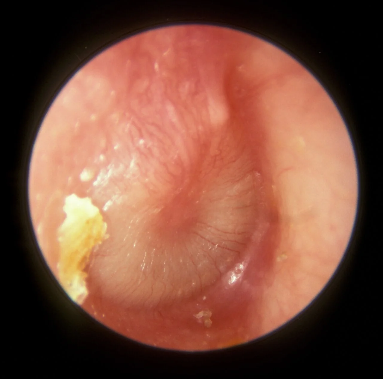
In OME the drum is typically dull, retracted, and grey or yellow, sometimes with visible air-fluid levels or bubbles, but without the erythema and bulging seen in acute infection. Pneumatic otoscopy, where available, shows reduced mobility of the drum in both conditions because the middle ear space is no longer air-filled.
Examine for tenderness or swelling over the mastoid process, which raises concern for mastoiditis, and check for facial nerve function, since the nerve runs through the middle ear and can rarely be affected by severe infection. Tympanometry, where available, is useful to confirm reduced compliance and support a diagnosis of effusion.
Differential diagnosis
Several conditions overlap with otitis media in presentation and should be considered, particularly when the picture is atypical or not settling as expected:1
- Otitis externa: pain worse on moving the pinna or tragus, normal or difficult-to-visualise drum, canal oedema
- Mastoiditis: post-auricular swelling, tenderness and erythema, protruding pinna
- Referred dental or temporomandibular pain: normal otoscopy
- Foreign body: especially in young children, with unilateral discharge or odour
- Bullous myringitis: haemorrhagic blisters on the drum, associated with Mycoplasma
- Cholesteatoma: chronic offensive discharge with an attic retraction pocket or crust, not an acute picture
Investigations
AOM is a clinical diagnosis made on history and otoscopy; investigations are not routinely required.1 Swabs of discharge are not useful in uncomplicated cases and do not change initial management.
If OME is suspected, audiometry and tympanometry are used to confirm a conductive hearing loss and reduced drum compliance, and to quantify severity before deciding on active observation versus referral. In children old enough to cooperate, pure tone audiometry is used; younger children may need play or visual reinforcement audiometry.2
Investigate further only if there is diagnostic doubt or a complication is suspected: FBC and CRP if the child is systemically unwell, and CT temporal bones or MRI if mastoiditis or an intracranial complication is being considered.
Management of acute otitis media
Most episodes of AOM resolve spontaneously within 3 days without antibiotics, because the underlying process is usually viral or a self-limiting bacterial infection.3 Analgesia with paracetamol or ibuprofen is the mainstay of initial management for every child, and parents should be given safety-netting advice about when to seek review.
Antibiotics are not routinely recommended for immediately-well children, but NICE NG91 identifies groups where an immediate prescription (or a backup prescription to use if not improving within 3 days) is appropriate:3
| Situation | Approach |
|---|---|
| Systemically well, symptoms <4 days, no high-risk features | No antibiotic, or a back-up prescription; analgesia and safety netting |
| Symptoms not improving after 3 days, or worsening | Offer an immediate antibiotic (or start the back-up prescription) |
| Otorrhoea from perforation | Offer an immediate antibiotic |
| Bilateral AOM in a child under 2 years | Offer an immediate antibiotic |
| Systemically unwell, or at risk of complications (e.g. significant comorbidity, immunocompromise, young infant) | Offer an immediate antibiotic and consider same-day assessment |
Amoxicillin for 5-7 days is first line; clarithromycin or erythromycin are alternatives in penicillin allergy. Co-amoxiclav is reserved for treatment failure or recurrent infection.4 Decongestants and antihistamines are not recommended, as they do not improve outcomes and add side effects.
Recurrent AOM (3 or more episodes in 6 months, or 4 or more in a year) warrants ENT referral to consider grommet insertion, and adenoidectomy is sometimes performed alongside if adenoidal hypertrophy is contributing to eustachian tube obstruction.
Management of otitis media with effusion
The majority of effusions resolve spontaneously, so the first step is a period of active observation for 6-12 weeks, with review and repeat audiometry to check for resolution.2 This avoids unnecessary surgery in the many children whose hearing recovers without intervention.
Referral for grommets (ventilation tubes) is considered if the effusion persists beyond 3 months, particularly if there is a persistent hearing loss of 25-30 dB or more in the better ear, or if hearing loss is affecting speech and language development, learning or behaviour. Adenoidectomy is often performed at the same time if adenoidal hypertrophy is contributing, and particularly in recurrent cases.2
Hearing aids are an alternative to surgery for children in whom surgery is not suitable or not wanted, and are the preferred option in children with Down syndrome or cleft palate, who have a higher rate of effusion recurrence after grommets.
Complications
Most children recover from AOM without any lasting problem, but complications do occur and understanding them explains why certain features prompt urgent referral rather than routine primary care management.1
- Tympanic membrane perforation: usually heals spontaneously within a few weeks; persistent perforation may need surgical repair (myringoplasty)
- Mastoiditis: infection spreading into the mastoid air cells, covered in detail separately
- Chronic suppurative otitis media: persistent otorrhoea through a non-healing perforation
- Hearing loss: usually conductive and temporary, but recurrent effusion can cause developmental and educational impact if unaddressed
- Labyrinthitis: spread of infection to the inner ear causing vertigo and sensorineural hearing loss
- Facial nerve palsy: rare, from nerve involvement as it passes through the middle ear
- Intracranial complications: meningitis, extradural or subdural abscess, and sigmoid sinus thrombosis are rare but serious
Red flags
Prognosis
AOM is a self-limiting illness in most children, with pain typically resolving within 3 days and the episode settling completely within a week. Around 80% of children improve within 3 days regardless of antibiotic use, which underlies the strategy of reserving antibiotics for those most likely to benefit.3
OME resolves spontaneously in the majority of children within 3 months, particularly effusions that follow an episode of AOM. A minority go on to have persistent or recurrent effusion requiring surgical intervention, and these children should be followed up to ensure hearing loss is not affecting their development.2
References
- NICE Clinical Knowledge Summaries (CKS). Otitis media - acute. 2023. Available here
- NICE Clinical Knowledge Summaries (CKS). Otitis media with effusion. 2022. Available here
- NICE NG91. Otitis media (acute): antimicrobial prescribing. 2022. Available here
- BNF. Amoxicillin - indications and dosing. Available here
- Michael Hawke MD, CC BY 4.0, via Wikimedia Commons. Available here
This article is written for revision and education. It is not clinical guidance and must not be used to make decisions about the care of a patient. Always check current NICE guidance and local protocols.