
Impetigo
Key points
- Impetigo: a highly contagious superficial bacterial skin infection caused by Staphylococcus aureus and/or Streptococcus pyogenes, most common in young children and spread readily by direct contact.
- Non-bullous impetigo: around 70% of cases, causing classic golden, honey-coloured crusted lesions, usually around the nose and mouth; caused by S. aureus alone or in combination with S. pyogenes.
- Bullous impetigo: caused exclusively by S. aureus strains producing exfoliative toxins that cleave desmoglein 1, causing flaccid, fluid-filled bullae that rupture to leave a thin, varnish-like brown crust.
- Contagiousness: spreads readily by direct contact and via fomites (towels, toys); outbreaks are common in nurseries and schools, and affected children should be excluded until lesions are crusted and healed, or for 48 hours after starting antibiotics.
- Diagnosis: clinical in most cases; a skin swab for culture and sensitivities is reserved for extensive, recurrent or treatment-resistant disease, or during a suspected outbreak.
- First-line treatment: hydrogen peroxide 1% cream for localised, non-bullous disease, reserving topical or oral antibiotics for more extensive or bullous disease, in order to reduce antibiotic resistance.
- Key complication: post-streptococcal glomerulonephritis can follow streptococcal impetigo and, unlike rheumatic fever, is not prevented by antibiotic treatment of the skin infection.
- Red flag: staphylococcal scalded skin syndrome - widespread, tender erythema with sheet-like skin detachment and a positive Nikolsky sign in a systemically unwell child - is a related but distinct dermatological emergency.
Introduction
Impetigo is a superficial, highly contagious bacterial infection of the epidermis, and one of the commonest skin infections seen in young children, with a peak incidence between 2 and 5 years old. It is caused by Staphylococcus aureus, Streptococcus pyogenes (group A streptococcus), or a combination of the two, and typically follows minor breaks in the skin barrier - a scratch, an insect bite, or eczema - that let bacteria establish infection in otherwise healthy skin.1
Two clinically distinct patterns exist - non-bullous and bullous - which differ in their mechanism, appearance and typical age group, though both are managed with the same broad principles: reduce transmission, and treat with antimicrobial therapy proportionate to the extent of disease rather than reaching for oral antibiotics as a default.
Pathophysiology
Non-bullous impetigo results from direct bacterial invasion of the superficial epidermis following a breach in the skin barrier. The organism - most often S. aureus, sometimes S. pyogenes, occasionally both together - proliferates within the epidermis and provokes a neutrophilic response, producing a vesicle or pustule that quickly ruptures. The exudate dries to form the characteristic golden-yellow crust, coloured by dried serum rather than pus.
Bullous impetigo is mechanistically different and is caused exclusively by S. aureus strains that produce exfoliative (epidermolytic) toxins A and B. These toxins are serine proteases that specifically cleave desmoglein 1, a desmosomal adhesion protein holding keratinocytes together in the superficial epidermis. Loss of desmoglein 1 function causes the superficial epidermis to split, producing a fragile, fluid-filled bulla that ruptures easily.6
Risk factors
- Young age - peak incidence in pre-school and primary-school-aged children
- Warm, humid weather - infection is commoner in summer months
- Close contact settings - nurseries, schools and households, given how readily impetigo spreads
- Pre-existing skin barrier disruption - atopic dermatitis, minor trauma, insect bites, scabies, and varicella lesions are all common portals of entry
- Poor hygiene and overcrowding
- Contact sports with skin-to-skin contact
- Nasal carriage of Staphylococcus aureus, which predisposes to recurrent episodes
Clinical features
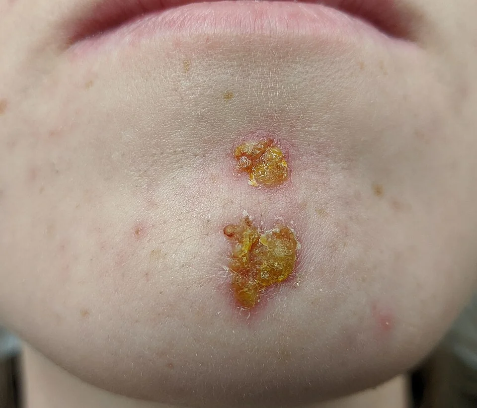
| Non-bullous | Bullous | |
|---|---|---|
| Proportion of cases | Around 70% | Around 30% |
| Organism | S. aureus alone, or with S. pyogenes | S. aureus only (exfoliative toxin-producing strains) |
| Typical age | Any age, commonly school-age children | Neonates and infants especially, though any age can be affected |
| Typical site | Around the nose and mouth, and any exposed or traumatised skin | Flexures, trunk and napkin area |
| Lesion | Vesicles or pustules that rupture quickly, forming a thick, golden-yellow crust | Flaccid, fragile bullae that rupture easily, leaving a thin, varnish-like brown crust and a collarette of scale |
| Systemic upset | Usually well, though regional lymphadenopathy can occur | Can be more unwell, particularly infants, with fever and irritability |
Lesions in both forms are typically mildly itchy or asymptomatic rather than painful, and multiple lesions often appear in clusters as the infection spreads locally through scratching or contact (autoinoculation).
Clinical examination
- Lesion morphology and distribution - crusted lesions around the nose/mouth versus flaccid bullae in flexures or the napkin area
- Extent - localised versus widespread disease, which determines whether topical or oral treatment is appropriate
- Regional lymphadenopathy
- Signs of a portal of entry - underlying eczema, an insect bite, or a scratch
- Systemic wellbeing - fever, lethargy or poor feeding in an infant should prompt consideration of more extensive staphylococcal disease, including SSSS
- Skin fragility and a positive Nikolsky sign (gentle lateral pressure causing the epidermis to shear) - if there is any suggestion of widespread skin involvement beyond the primary lesions
Differential diagnosis
- Herpes simplex (cold sores) - grouped, monomorphic vesicles on an erythematous base, often recurrent at the same site, rather than the honey-crusted plaques of impetigo
- Eczema herpeticum - punched-out vesicles and erosions on a background of atopic dermatitis with systemic upset, a dermatological emergency needing aciclovir rather than antibiotics alone
- Cellulitis - deeper, more diffuse erythema, warmth and tenderness, usually unilateral, without the superficial crusting of impetigo
- Tinea corporis (ringworm) - an annular, expanding plaque with an active scaly edge rather than a honey-coloured crust
- Contact or atopic dermatitis - can look superficially similar, especially if secondarily infected, but lacks the primary vesicopustular stage
- Bullous pemphigoid or pemphigus - autoimmune blistering diseases to consider if bullae are tense (pemphigoid) or very fragile with oral involvement (pemphigus) and not responding to antimicrobial treatment, particularly in an adult
- Varicella (chickenpox) - vesicles at different stages across the whole body rather than clustered at one or two sites, usually with a preceding prodrome
Investigations
Impetigo is usually a clinical diagnosis, and most cases do not need any investigation before starting treatment.
- Skin swab for bacterial culture and sensitivities - reserved for extensive disease, recurrent impetigo, disease not responding to first-line treatment, or a suspected outbreak, and useful for identifying MRSA where relevant
- Nasal swab - considered in recurrent impetigo to identify staphylococcal carriage, which can then be treated to reduce reinfection
- Urinalysis - checked a few weeks after streptococcal impetigo if there are any symptoms suggesting post-streptococcal glomerulonephritis (haematuria, oedema, hypertension), though routine screening of asymptomatic patients is not recommended
Management
Management follows a stepwise approach based on extent, chosen partly to reduce unnecessary antibiotic use and resistance.2
- Hygiene advice for everyone - avoid sharing towels, flannels and bedding; wash affected areas gently with soap and water; keep nails short to reduce spread by scratching
- School or nursery exclusion - until lesions are crusted and healed, or for 48 hours after starting an effective antibiotic, whichever is sooner4
- Localised, non-bullous impetigo - hydrogen peroxide 1% cream is first-line, as effective as topical antibiotics for limited disease with a lower risk of promoting antibiotic resistance
- Topical fusidic acid - an alternative first-line option, or used where hydrogen peroxide is not suitable or has failed; short courses are preferred to reduce the risk of resistance developing, which is a recognised problem with prolonged fusidic acid use3
- Widespread non-bullous impetigo, or any bullous impetigo - a short course of oral flucloxacillin (or oral erythromycin/clarithromycin in penicillin allergy)
- Extensive, systemically unwell, or immunocompromised patients - may need admission and intravenous antibiotics
- Recurrent impetigo - consider a nasal decolonisation regimen (topical nasal mupirocin) if staphylococcal carriage is confirmed, alongside review of any underlying skin condition such as eczema that is providing repeated portals of entry
Complications
- Post-streptococcal glomerulonephritis - can follow streptococcal skin or throat infection; unlike rheumatic fever, treating the preceding impetigo does not reliably prevent it, since the nephritogenic immune response is already established by the time skin infection is recognised7
- Staphylococcal scalded skin syndrome - widespread toxin-mediated skin detachment at a site distant from the original infection, discussed under Red flags
- Cellulitis - if infection extends into the deeper dermis and subcutaneous tissue
- Scarring - unusual with impetigo itself, since it is a superficial infection, but can occur if secondarily excoriated or infected more deeply
- Recurrent infection - particularly where nasal or perineal staphylococcal carriage, or an underlying skin condition, is not addressed
- Rarely, invasive streptococcal or staphylococcal disease - bacteraemia or toxic shock syndrome, exceptionally uncommon but recognised, particularly in extensive or neglected infection
Red flags
Prognosis
Impetigo is generally a mild, self-limiting infection that responds well to appropriate topical or oral treatment, typically clearing within 7-10 days. Even untreated, most cases resolve within a few weeks, though treatment shortens the infectious period and reduces the risk of spread to others.5
The rare but important complications - post-streptococcal glomerulonephritis and staphylococcal scalded skin syndrome - are the main determinants of a poor outcome, and neither is reliably prevented by prompt antibiotic treatment of the skin lesions themselves. This is worth explaining clearly to parents: treating the visible infection promptly reduces spread and speeds resolution, but does not eliminate every possible complication, and they should be advised to seek review if the child becomes systemically unwell or develops new symptoms such as dark urine or joint pains in the following weeks.
References
- NICE Clinical Knowledge Summaries. Impetigo. Available here
- NICE NG153. Impetigo: antimicrobial prescribing. 2020. Available here
- BNF. Fusidic acid. Available here
- UK Health Security Agency. Guidance on infection control in schools and other childcare settings. Available here
- Hartman-Adams H, Banvard C, Juckett G. Impetigo: diagnosis and treatment. American Family Physician. 2014. Available here
- Amagai M, Matsuyoshi N, Wang ZH et al. Toxin in bullous impetigo and staphylococcal scalded-skin syndrome targets desmoglein 1. Nature Medicine. 2000. Available here
- Rodriguez-Iturbe B, Musser JM. The current state of poststreptococcal glomerulonephritis. Journal of the American Society of Nephrology. 2008. Available here
- Handler MZ, Schwartz RA. Staphylococcal scalded skin syndrome: diagnosis and management in children and adults. Journal of the European Academy of Dermatology and Venereology. 2014. Available here
This article is written for revision and education. It is not clinical guidance and must not be used to make decisions about the care of a patient. Always check current NICE guidance and local protocols.