
Cutaneous Warts
Key points
- Cutaneous warts: benign epidermal proliferations caused by human papillomavirus (HPV) infecting keratinocytes, with the clinical appearance and behaviour depending on the infecting HPV subtype and the body site.
- Common types: common warts (verruca vulgaris), plantar warts (verruca plantaris, including the deep, painful myrmecia), plane warts (verruca plana), and filiform warts.
- Transmission: direct skin contact or contact with contaminated surfaces (communal showers, swimming pools), inoculating the virus through minor breaks in the skin barrier; incubation is weeks to months.
- The paring sign: shaving the surface of a wart reveals pinpoint bleeding from thrombosed capillaries and interruption of normal skin lines - the key clinical feature distinguishing a wart from a corn or callus.
- Natural history: the great majority resolve spontaneously within 1-2 years, especially in children, as cell-mediated immunity clears the infection - a fact that should guide the decision of whether to treat at all.
- First-line treatment: topical salicylic acid, with or without paring, is at least as effective as cryotherapy and is preferred for its lower cost and lack of pain; cryotherapy is added for resistant lesions.
- Immunosuppression: warts are more numerous, persistent and treatment-resistant in immunosuppressed patients, particularly organ transplant recipients, who also carry a raised long-term risk of cutaneous squamous cell carcinoma.
- Red flag: a rapidly growing, bleeding, or atypical 'wart', especially in an immunosuppressed or elderly patient, should be biopsied to exclude squamous cell carcinoma or keratoacanthoma masquerading as a wart.
Introduction
Cutaneous warts are extremely common, affecting an estimated 10% of children and young adults at any one time, with the highest incidence between ages 12 and 16. They are caused by infection of keratinocytes with human papillomavirus (HPV), a large family of viruses with over 200 identified subtypes, of which a relatively small number cause skin (as opposed to anogenital or mucosal) warts.1,2
Most cutaneous warts are a nuisance rather than a medical problem, and the single most useful piece of management advice is also the least prescribed: since most warts clear on their own, watchful waiting is a completely reasonable first option, particularly in children. This note focuses on cutaneous warts; anogenital warts, caused by different HPV subtypes and raising different considerations including sexual health screening and safeguarding in children, are a distinct topic.
Aetiology and pathophysiology
HPV infects basal keratinocytes through small breaks in the epidermis and drives their proliferation, producing the thickened, hyperkeratotic epidermal growth seen clinically. Different HPV subtypes have a preference for particular body sites and produce recognisably different clinical patterns, which is why 'a wart' is better thought of as a family of related presentations rather than one single disease.
- HPV 1, 2, 4, 27, 57 - common and plantar warts
- HPV 3, 10 - plane (flat) warts
- HPV 6, 11 - anogenital warts (and some benign laryngeal papillomas), covered separately
- HPV 16, 18 and other high-risk mucosal subtypes - genital and cervical dysplasia and malignancy, not associated with ordinary cutaneous warts
The virus remains confined to the epidermis and does not enter the systemic circulation, which is why cutaneous warts, however extensive, are a local skin problem rather than a systemic infection. Clearance depends on an effective cell-mediated immune response, which explains both why warts are so much more persistent in immunosuppressed patients, and why most eventually resolve spontaneously as the immune system 'catches up' with the infection.
Risk factors
- Childhood and adolescence - the age group with the highest prevalence, reflecting both exposure and an immune response that has not yet cleared the virus
- Immunosuppression - organ transplant recipients, HIV infection, and other causes of impaired cell-mediated immunity are associated with more numerous, larger and more treatment-resistant warts8
- Communal wet areas - swimming pools, communal showers and changing rooms, particularly relevant to plantar warts
- Occupational exposure - meat, poultry and fish handlers have a recognised excess of hand warts ('butcher's warts', associated with HPV 7)
- Nail biting and periungual skin picking - creates a portal of entry around the nail folds
- Eczema and other conditions that disrupt the skin barrier
- Direct contact with a household member or close contact with warts
Clinical features
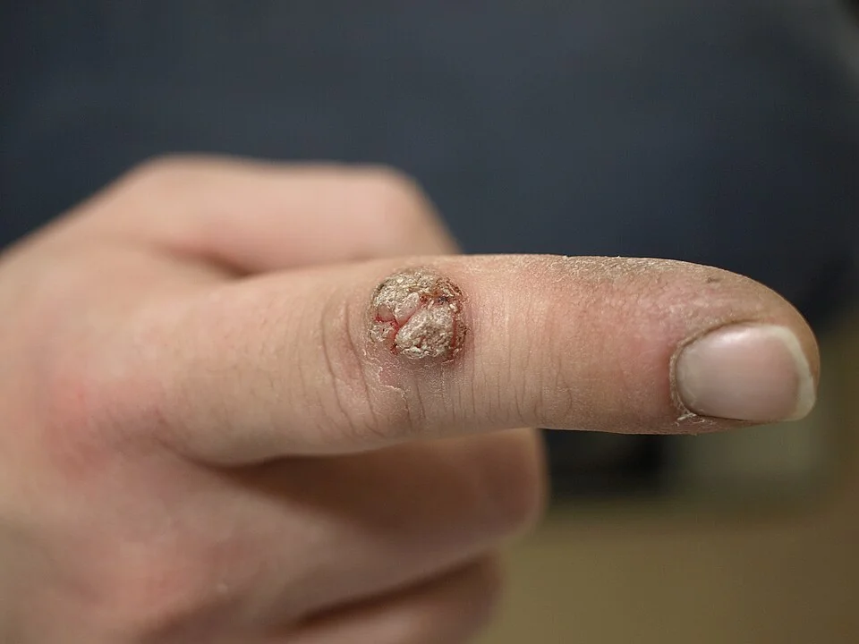
| Type | Features |
|---|---|
| Common wart (verruca vulgaris) | Firm, rough, hyperkeratotic, papillomatous papule, most often on the hands and fingers, especially around the nail folds |
| Plantar wart (verruca plantaris) | Flattened by pressure into the sole rather than raised; can be painful on weight-bearing |
| Myrmecia | A deep, painful, well-defined plantar wart caused by specific HPV subtypes, extending deeper into the dermis than typical plantar warts |
| Mosaic wart | Multiple small plantar warts coalescing into a larger plaque, usually less painful than a solitary deep lesion |
| Plane wart (verruca plana) | Small, smooth-surfaced, flat-topped papules, often numerous, commonly on the face and dorsum of the hands; can spread along scratch lines (Koebner phenomenon) |
| Filiform wart | A thin, finger-like projection, typically on the face or neck |
Regardless of type, close inspection typically shows an interruption of the normal skin lines (dermatoglyphics) across the surface of the lesion, and paring the surface reveals pinpoint bleeding points from thrombosed dermal capillaries - together the most useful bedside features for confirming the diagnosis.
Clinical examination
- Site, number and distribution - solitary versus multiple lesions, and characteristic sites (periungual, plantar, facial)
- Surface features - interruption of skin lines and pinpoint bleeding on paring
- Tenderness on pressure - particularly relevant for plantar warts, which can affect gait
- Signs of immunosuppression or an underlying cause if warts are unusually extensive, numerous or resistant to treatment
- Any rapidly growing, ulcerating or atypical lesion, which should prompt reconsideration of the diagnosis rather than assumption of a resistant wart
Differential diagnosis
- Corn or callus - preserves normal skin lines, no bleeding points on paring, related to focal pressure
- Seborrhoeic keratosis - a stuck-on, warty-looking lesion but typically in older adults, often pigmented, and without the bleeding points of a true wart
- Molluscum contagiosum - a different virus (a poxvirus) producing dome-shaped, umbilicated papules rather than a rough, papillomatous surface
- Squamous cell carcinoma or keratoacanthoma - can mimic a wart, particularly on the hands or in sun-damaged or immunosuppressed skin, and should be considered in any rapidly growing or atypical lesion, especially in an older or immunosuppressed patient
- Amelanotic or acral lentiginous melanoma - a rare but important differential for an atypical plantar lesion that does not behave like a typical wart
- Digital myxoid (mucous) cyst - a smooth, translucent, fluid-filled swelling near a distal interphalangeal joint, unlike the rough surface of a wart
Investigations
Cutaneous warts are diagnosed clinically, and investigation is reserved for atypical or diagnostically uncertain lesions.
- Dermoscopy - can help confirm the diagnosis by showing the characteristic thrombosed capillary loops and interrupted skin lines, and helps distinguish a wart from a corn or an early acral melanoma
- Skin biopsy - reserved for lesions that are rapidly growing, ulcerating, bleeding spontaneously, or otherwise atypical, particularly in an immunosuppressed or elderly patient, to exclude squamous cell carcinoma or keratoacanthoma
Management
Because most cutaneous warts eventually resolve on their own, the first management decision is whether treatment is needed at all - not every wart requires active intervention.3
- No treatment (watchful waiting) - a reasonable choice, especially in children, for asymptomatic warts, since spontaneous resolution within 1-2 years is the norm
- Topical salicylic acid, applied regularly after paring or soaking the wart, is first-line active treatment and is at least as effective as cryotherapy in trials, with less discomfort and lower cost4,5
- Cryotherapy (liquid nitrogen) - added or used instead where topical treatment fails, typically repeated every 2-3 weeks; more painful and can cause blistering or, rarely, scarring or pigment change
- Duct tape occlusion - a simple, low-cost adjunct with mixed evidence, sometimes used alongside paring
- Second-line options for resistant warts - topical immunotherapy (diphenylcyclopropenone), intralesional immunotherapy or bleomycin, and laser or surgical treatment, generally under specialist care given the higher risk of scarring6
In immunosuppressed patients, warts are often more numerous and considerably more resistant to standard treatment, and a lower threshold for specialist dermatology referral is appropriate, both to manage the warts themselves and because of the associated long-term skin cancer risk in this group.
Complications
- Pain and functional impact - particularly deep plantar warts (myrmecia) affecting gait
- Autoinoculation and spread - to adjacent skin or other body sites through scratching or shaving
- Scarring - from aggressive or repeated cryotherapy, or from surgical removal, which is one reason destructive treatment is reserved for resistant lesions rather than used first-line
- Psychological and social impact - particularly visible facial or hand warts in children and adolescents
- Persistent extensive warts in immunosuppressed patients, with an associated long-term increase in cutaneous squamous cell carcinoma risk, particularly in organ transplant recipients on long-term immunosuppression8
Red flags
Prognosis
The natural history of cutaneous warts is genuinely favourable: in immunocompetent individuals, particularly children, roughly two-thirds resolve spontaneously within 2 years without any treatment, as cell-mediated immunity clears the infection.7
Treatment shortens this course and reduces spread but does not reliably prevent recurrence, and even successfully treated warts can recur from residual virus in adjacent, clinically normal skin. The main exception to this generally reassuring picture is the immunosuppressed patient, in whom warts are frequently extensive, persistent despite treatment, and a marker of a population that also needs long-term skin cancer surveillance.
References
- NICE Clinical Knowledge Summaries. Warts and verrucae. Available here
- British Association of Dermatologists. Viral warts patient information leaflet. Available here
- Sterling JC, Gibbs S, Haque Hussain SS et al. British Association of Dermatologists' guidelines for the management of cutaneous warts 2014. Available here
- Kwok CS, Gibbs S, Bennett C et al. Topical treatments for cutaneous warts. Cochrane Database of Systematic Reviews. 2012. Available here
- Bruggink SC, Gussekloo J, Berger MY et al. Cryotherapy with liquid nitrogen versus topical salicylic acid application for cutaneous warts in primary care. CMAJ. 2010. Available here
- Bacelieri R, Johnson SM. Cutaneous warts: an evidence-based approach to therapy. American Family Physician. 2005. Available here
- DermNet NZ. Viral warts. Available here
- Euvrard S, Kanitakis J, Claudy A. Skin cancers after organ transplantation. New England Journal of Medicine. 2003. Available here
This article is written for revision and education. It is not clinical guidance and must not be used to make decisions about the care of a patient. Always check current NICE guidance and local protocols.