
Cutaneous Fungal Infection
Key points
- Dermatophytes: a group of fungi (Trichophyton, Microsporum, Epidermophyton species) that digest keratin, confining infection to the stratum corneum, hair and nails - collectively called tinea or ringworm.
- Naming convention: tinea infections are named by body site using Latin - corporis (body), cruris (groin), pedis (foot), capitis (scalp), unguium (nail), manuum (hand).
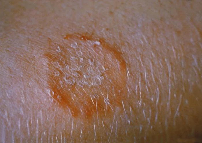
- Classic appearance: an expanding, annular, erythematous plaque with a raised, scaly, active edge and central clearing - the origin of the name 'ringworm', despite no worm being involved.
- Pityriasis versicolor: caused by the commensal yeast Malassezia, not a dermatophyte, producing hypo- or hyperpigmented, finely scaly macules on the trunk, most noticeable after sun exposure.
- Diagnosis: usually clinical, confirmed where needed with skin scrapings, nail clippings or plucked hairs sent for microscopy (potassium hydroxide preparation) and fungal culture.
- Topical treatment: sufficient for most localised skin infections - terbinafine or an azole cream applied well beyond the visible edge and continued for 1-2 weeks after clearance.
- Oral treatment needed: for tinea capitis, tinea unguium (onychomycosis), and any widespread, resistant or immunocompromised disease, since topical treatment cannot penetrate hair follicles or nail plates adequately.
- Tinea incognito: a dermatophyte infection modified and worsened by inappropriate topical corticosteroid treatment, losing its classic scaly, well-demarcated appearance and becoming harder to recognise.
Introduction
Fungal skin infections are extremely common and fall into two quite different groups: infections with dermatophytes, a specialised group of fungi that digest keratin and are collectively called tinea or ringworm, and infections with yeasts, chiefly Malassezia (pityriasis versicolor) and Candida (intertrigo and mucocutaneous candidiasis).
Despite the name, ringworm has nothing to do with worms - the term describes the characteristic ring-shaped, expanding plaque that dermatophyte infection produces on smooth skin. Recognising the pattern, naming the site correctly, and knowing when topical treatment is enough versus when oral therapy is required are the practical skills this topic tests most often.
Aetiology and classification
Three genera of dermatophyte cause the great majority of human infection: Trichophyton, Microsporum and Epidermophyton. All three secrete keratinases that allow them to digest keratin, which is why infection is confined to the dead, keratinised layers of the epidermis, hair and nails and never invades living tissue - dermatophytes do not cause systemic infection in an immunocompetent host.
- Anthropophilic species (person-to-person spread) - the commonest source in the UK, spread by direct contact or shared items such as towels and combs
- Zoophilic species (animal-to-person spread) - from cats, dogs, or farm animals, and tend to produce a more intensely inflammatory reaction in humans
- Geophilic species (soil-to-person spread) - less common, acquired from contaminated soil
Tinea is named by combining 'tinea' with the Latin term for the affected site, which is worth learning as a fixed list since exam questions rely on precise terminology.
| Term | Site |
|---|---|
| Tinea corporis | Trunk and limbs (body) |
| Tinea cruris | Groin |
| Tinea pedis | Feet ('athlete's foot') |
| Tinea manuum | Hands |
| Tinea capitis | Scalp |
| Tinea unguium (onychomycosis) | Nails |
| Tinea faciei | Face |
Pityriasis versicolor is a separate entity caused by Malassezia species, a lipophilic yeast that is part of normal skin flora. Under certain conditions - heat, humidity, sebaceous skin, immunosuppression - it converts to its pathogenic hyphal form and produces the characteristic scaly, discoloured macules, particularly on the trunk.
Risk factors
- Warm, humid environments and occlusive footwear or clothing, predisposing to tinea pedis and tinea cruris
- Communal facilities - swimming pools, showers and changing rooms, particularly for tinea pedis
- Close contact with infected people, pets or farm animals
- Contact sports with skin-to-skin contact, classically tinea corporis in wrestlers (tinea gladiatorum)
- Immunosuppression (diabetes, HIV, immunosuppressive drugs) - predisposes to more extensive, atypical or treatment-resistant infection
- Peripheral vascular disease and diabetes - particularly relevant to tinea pedis and onychomycosis, and to the risk of secondary bacterial infection
- Hyperhidrosis
Clinical features
The classic dermatophyte lesion on smooth skin is an annular, erythematous, expanding plaque with a raised, scaly, active edge and relative central clearing - the pattern that gives ringworm its name.
| Site | Features |
|---|---|
| Tinea corporis | One or more annular plaques with an active scaly edge, on the trunk or limbs |
| Tinea cruris | Well-demarcated, itchy erythema in the groin, extending onto the upper thigh; typically spares the scrotum, which is a useful clue against candidal intertrigo |
| Tinea pedis | Itchy, macerated scaling in the toe web spaces (most often), or a diffuse 'moccasin' scaly pattern over the sole and sides of the foot |
| Tinea capitis | Patchy hair loss with scaling; can progress to broken-off hair stumps ('black dot' pattern), diffuse fine scale, or a boggy, pustular, inflamed swelling (kerion) |
| Tinea unguium (onychomycosis) | Thickened, discoloured (yellow-white), crumbly nail, usually starting distally and spreading proximally; toenails more often affected than fingernails |
| Tinea manuum | Fine scaling, often unilateral, frequently accompanying tinea pedis on the same side ('two feet, one hand' pattern) |
Pityriasis versicolor
Presents as multiple round or oval macules with fine surface scale, mainly on the upper trunk, neck and upper arms. Lesions can be hypopigmented on tanned or darker skin, or pink-to-brown on paler skin - hence 'versicolor' - and become more noticeable in summer as surrounding skin tans while affected areas do not.
Clinical examination
- Morphology and margin - an active, scaly, raised edge with central clearing is the key feature to look for on smooth skin
- Distribution - single versus multiple lesions, and specifically the toe webs, groin and scalp
- Hair and scalp - patchy alopecia, scaling, broken hair stumps, or a boggy inflamed swelling (kerion)
- Nails - thickening, discolouration and subungual debris, and whether one or multiple nails are affected
- Regional lymphadenopathy - can accompany more inflammatory zoophilic infections or a kerion
Differential diagnosis
- Nummular (discoid) eczema - less well demarcated, without the same active scaly edge, and usually more symmetrical
- Psoriasis - well-demarcated plaques with silvery scale, but static rather than expanding, and typically affecting extensor surfaces and the scalp margin symmetrically
- Pityriasis rosea - an initial herald patch followed by a 'Christmas tree' distribution of smaller oval lesions on the trunk
- Candidal intertrigo - affects flexures and the scrotum (unlike tinea cruris, which tends to spare it), with satellite pustules at the margin
- Erythrasma - a bacterial (Corynebacterium minutissimum) intertriginous infection that fluoresces coral-pink under Wood's lamp, unlike tinea
- Alopecia areata - smooth, non-scaly patches of hair loss without scale, unlike tinea capitis
Investigations
Localised, classic-looking dermatophyte infection on smooth skin can often be diagnosed and treated clinically. Confirmation is more important before committing to prolonged oral treatment, as for the scalp or nails, or where the diagnosis is uncertain.7
- Skin scrapings from the active edge, or plucked hairs, or nail clippings including subungual debris, sent for microscopy with a potassium hydroxide (KOH) preparation - shows branching hyphae
- Fungal culture - takes several weeks but identifies the specific organism, useful before starting oral treatment for tinea capitis or onychomycosis, and helpful in guiding public health advice if a zoophilic species is found
- Wood's lamp examination - some but not all Microsporum species fluoresce green under UV light; a negative result does not exclude tinea capitis, since Trichophyton tonsurans (the commonest UK cause) does not fluoresce
- Wood's lamp for pityriasis versicolor - characteristically shows a yellow-gold fluorescence
Management
Localised skin, groin and foot infection
- Topical terbinafine or an azole cream (clotrimazole, miconazole) applied to the lesion and at least 1-2 cm beyond its visible edge, continued for 1-2 weeks after the rash has cleared to reduce relapse1,2
- Keep the area dry, wear breathable footwear and change socks daily for tinea pedis, and avoid sharing towels
- Oral terbinafine or itraconazole for widespread disease, infection resistant to topical treatment, or immunosuppressed patients
Scalp infection (tinea capitis)
Always requires oral antifungal treatment - typically griseofulvin or terbinafine - because topical treatment cannot penetrate the hair follicle to reach the fungus. Topical antifungal shampoo (for example ketoconazole) is used alongside oral treatment to reduce spore shedding and transmission to others while the oral course takes effect.8
Nail infection (onychomycosis)
Oral terbinafine is first-line for confirmed dermatophyte nail infection, typically for 6 weeks (fingernails) to 3 months (toenails), since topical treatment alone rarely clears established nail disease. Baseline liver function is checked before starting, given the (rare) risk of hepatotoxicity. Topical nail lacquer (amorolfine) is an option for very early, limited disease or where oral treatment is contraindicated, though cure rates are lower.3,5
Pityriasis versicolor
Topical ketoconazole shampoo used as a wash, or a topical azole cream for limited areas, is first-line. Widespread or recurrent disease can be treated with a short course of oral itraconazole. Patients should be warned that pigment changes can take several months to resolve even after the yeast has been successfully treated, since it reflects a temporary disturbance of melanocyte function rather than ongoing infection.6
Complications
- Tinea incognito - a masked, spreading infection following inappropriate topical corticosteroid use
- Kerion and scarring alopecia - from an intensely inflammatory, untreated or delayed-treatment tinea capitis
- Secondary bacterial infection - particularly in macerated tinea pedis, which can be a portal of entry for cellulitis, especially in patients with diabetes or lymphoedema
- Id reaction - a distant, itchy, vesicular eruption (often on the hands) representing an immune reaction to the fungal infection elsewhere, rather than a separate infection needing its own antifungal treatment
- Permanent nail damage - if onychomycosis is longstanding and untreated
Red flags
Prognosis
Localised dermatophyte infections of smooth skin, the groin and the feet generally clear well with appropriate topical treatment within 2-4 weeks, though recurrence is common if the predisposing environment (damp footwear, shared facilities, an infected household contact or pet) is not addressed.4
Scalp and nail infections take considerably longer to clear because of the months-long growth cycle of hair and nail, and onychomycosis in particular has a meaningful relapse rate even after apparently successful oral treatment. Pityriasis versicolor responds well to treatment, but pigment change can persist for months, and recurrence is common because the causative yeast is a normal skin commensal that simply reactivates under the right conditions rather than being an infection to permanently eradicate.
References
- NICE Clinical Knowledge Summaries. Fungal skin infection - body and groin. Available here
- NICE Clinical Knowledge Summaries. Fungal skin infection - foot. Available here
- NICE Clinical Knowledge Summaries. Fungal nail infection. Available here
- NICE Clinical Knowledge Summaries. Fungal skin infection - scalp. Available here
- BNF. Fungal infections - treatment summary. Available here
- DermNet NZ. Pityriasis versicolor. Available here
- Ely JW, Rosenfeld S, Seabury Stone M. Diagnosis and management of tinea infections. American Family Physician. 2014. Available here
- Gupta AK, Mays RR, Versteeg SG et al. Tinea capitis in children: a systematic review of management. Journal of the European Academy of Dermatology and Venereology. 2018. Available here
This article is written for revision and education. It is not clinical guidance and must not be used to make decisions about the care of a patient. Always check current NICE guidance and local protocols.