
Chronic Heart Failure: Diagnosis and Management
Key points
- Chronic heart failure: a clinical syndrome of breathlessness, fatigue and fluid retention caused by a structural or functional cardiac abnormality.
- Classification: by ejection fraction - reduced (HFrEF, LVEF 40% or below) or preserved (HFpEF, LVEF 50% or above). The treatments differ substantially.
- Commonest cause: ischaemic heart disease, followed by hypertension, valve disease and cardiomyopathy.
- Diagnosis: measure NT-proBNP first. Above 2000 ng/L needs specialist assessment and echocardiography within 2 weeks; 400-2000 ng/L within 6 weeks.
- Symptoms: breathlessness, orthopnoea, paroxysmal nocturnal dyspnoea, fatigue and ankle swelling, graded by NYHA class.
- First-line drugs: an ACE inhibitor and a beta-blocker, started one at a time and titrated slowly.
- The four pillars: ACE inhibitor or ARNI, beta-blocker, mineralocorticoid receptor antagonist and SGLT2 inhibitor all reduce mortality in HFrEF.
- Diuretics: relieve congestion and improve symptoms but do not improve survival - use the lowest dose that keeps the patient dry.
Introduction
Chronic heart failure is a clinical syndrome, not a diagnosis in itself. It describes the symptoms and signs that result when a structural or functional abnormality of the heart reduces cardiac output, raises intracardiac pressures, or both.1
It affects roughly 1 million people in the UK, with prevalence rising steeply with age - over 10% of people above 80. It accounts for around 5% of emergency medical admissions and is one of the commonest reasons for readmission.2
The single most important distinction is by ejection fraction, because it determines whether the patient benefits from the drugs that improve survival.
| Type | LVEF | Notes |
|---|---|---|
| Reduced ejection fraction (HFrEF) | 40% or below | Systolic failure. The type with strong evidence for disease-modifying drugs. |
| Mildly reduced (HFmrEF) | 41-49% | An intermediate group; increasingly treated like HFrEF. |
| Preserved ejection fraction (HFpEF) | 50% or above | Diastolic failure - a stiff ventricle that fills poorly. Common in older, hypertensive and diabetic patients. |
Aetiology
Any process that damages the myocardium, overloads it, or prevents it filling can cause heart failure.
| Mechanism | Causes |
|---|---|
| Myocardial damage | Ischaemic heart disease (the commonest cause in the UK), myocarditis, cardiotoxic drugs |
| Pressure overload | Hypertension, aortic stenosis, coarctation, pulmonary hypertension (right heart) |
| Volume overload | Mitral or aortic regurgitation, ventricular septal defect |
| Cardiomyopathy | Dilated, hypertrophic, restrictive; alcohol; peripartum; genetic; amyloidosis, haemochromatosis, sarcoidosis |
| Arrhythmia | Atrial fibrillation, and tachycardia-induced cardiomyopathy from any sustained tachyarrhythmia |
| Impaired filling | Constrictive pericarditis, cardiac tamponade, restrictive cardiomyopathy |
| High output states | Severe anaemia, thyrotoxicosis, Paget disease, arteriovenous fistula, beriberi (thiamine deficiency) |
Why the drugs work
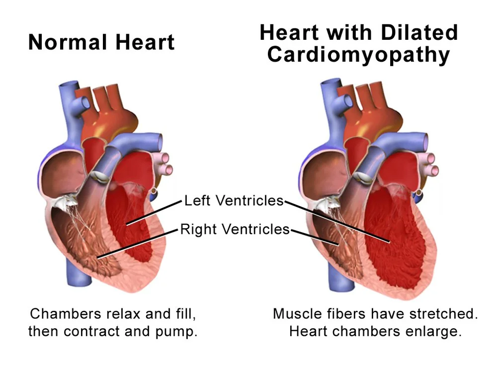
Falling cardiac output triggers compensatory neurohormonal activation: the renin-angiotensin-aldosterone system and the sympathetic nervous system. In the short term this maintains blood pressure and perfusion. Sustained, it is destructive - causing vasoconstriction, sodium and water retention, and progressive ventricular dilatation and fibrosis known as remodelling.
This is the key insight in heart failure pharmacology. ACE inhibitors, beta-blockers and mineralocorticoid receptor antagonists all improve survival because they interrupt this maladaptive response. Diuretics relieve congestion but do nothing to it, which is why they improve symptoms without improving survival.
Clinical features
Symptoms arise from congestion behind the failing ventricle and from inadequate output in front of it.
| Left heart failure | Right heart failure |
|---|---|
| Breathlessness on exertion | Ankle and leg swelling |
| Orthopnoea | Abdominal distension and ascites |
| Paroxysmal nocturnal dyspnoea | Nausea and early satiety from hepatic congestion |
| Fatigue and reduced exercise tolerance | Right upper quadrant discomfort |
| Nocturnal cough | Weight gain from fluid |
In practice most patients have congestive cardiac failure, with features of both - left heart failure raises pulmonary pressures, which in time causes the right ventricle to fail as well.
Grading severity
The New York Heart Association (NYHA) classification grades functional limitation and should be recorded at every review, since change over time guides treatment.3
| Class | Limitation |
|---|---|
| I | No limitation. Ordinary activity causes no symptoms. |
| II | Slight limitation. Comfortable at rest; ordinary activity causes symptoms. |
| III | Marked limitation. Comfortable at rest; less than ordinary activity causes symptoms. |
| IV | Symptoms at rest, worsened by any activity. |
Points to cover in the history
- Exercise tolerance in concrete terms, and whether it has changed
- Number of pillows, and any waking at night breathless
- Weight trend - a rapid gain means fluid, not fat
- Full cardiac history: previous myocardial infarction, hypertension, valve disease, arrhythmia
- Alcohol intake, and any chemotherapy or radiotherapy
- Current medications, including anything that worsens heart failure - NSAIDs, corticosteroids, verapamil, pioglitazone
- Adherence, and understanding of fluid and salt restriction
Clinical examination
Examination assesses fluid status, the severity of impairment, and the underlying cause.
- General: breathlessness at rest, cachexia in advanced disease, cyanosis
- Pulse: rate and rhythm - atrial fibrillation is both cause and consequence; a low-volume pulse and pulsus alternans in severe impairment
- JVP: raised, and one of the most useful signs of fluid overload
- Praecordium: displaced, diffuse apex beat indicating cardiomegaly; a third heart sound producing a gallop rhythm; murmurs of mitral or aortic disease
- Chest: bibasal fine inspiratory crackles; dullness at the bases from pleural effusions
- Abdomen: tender smooth hepatomegaly from congestion, ascites
- Legs: pitting oedema, which in a patient who has been in bed collects over the sacrum rather than the ankles
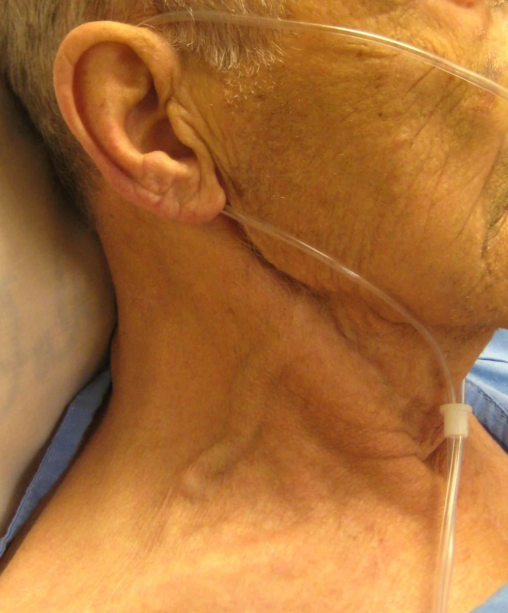
The most useful bedside measures of congestion are the JVP and the daily weight. Peripheral oedema alone is a poor guide - it has many other causes, including calcium channel blockers, venous insufficiency, hypoalbuminaemia and immobility.
Investigations
Natriuretic peptides decide the pathway
NICE recommends measuring NT-proBNP in anyone with suspected chronic heart failure. The result determines how quickly they need an echocardiogram, and it is a very common exam question.1
| NT-proBNP | Action |
|---|---|
| Above 2000 ng/L | Refer urgently - specialist assessment and echocardiography within 2 weeks |
| 400 to 2000 ng/L | Refer - specialist assessment and echocardiography within 6 weeks |
| Below 400 ng/L | Heart failure is less likely; consider an alternative diagnosis |
Echocardiography
Transthoracic echocardiography is the definitive test. It measures ejection fraction, which determines treatment; identifies the cause - regional wall motion abnormalities suggest ischaemia, valve lesions, hypertrophy; and assesses diastolic function, chamber sizes and pulmonary pressures.
Supporting investigations
- ECG - rarely normal in heart failure. Look for prior infarction, left ventricular hypertrophy, atrial fibrillation, and left bundle branch block, which is relevant to cardiac resynchronisation therapy.
- Chest X-ray - cardiomegaly, pulmonary congestion, effusions; also excludes other causes of breathlessness
- Bloods - FBC (anaemia), U&Es (baseline before ACE inhibitors and MRAs), LFTs, TFTs, HbA1c, lipids, and iron studies, since iron deficiency is common and treating it improves symptoms even without anaemia
- Spirometry - to identify coexisting COPD, which frequently confuses the picture
- Coronary angiography or CT coronary angiography - where ischaemia is the suspected cause and revascularisation is being considered
Management
Management has three aims: relieve symptoms, slow disease progression, and reduce mortality. Which drugs achieve the last of these depends entirely on the ejection fraction.
All patients
- Supervised exercise-based cardiac rehabilitation - improves symptoms, quality of life and hospital admission rates, and is recommended for anyone who is stable
- Annual influenza vaccination and one-off pneumococcal vaccination
- Smoking cessation and alcohol reduction, with abstinence where alcohol is the cause
- Salt and fluid advice - avoid salt substitutes containing potassium, which are dangerous alongside ACE inhibitors and MRAs
- Daily weights with a written plan on what to do if weight rises
- Review of drugs that worsen heart failure - NSAIDs, corticosteroids, verapamil and diltiazem, pioglitazone
- Specialist heart failure multidisciplinary team input, including a heart failure nurse
HFrEF: the drugs that improve survival
NICE first-line therapy is an ACE inhibitor and a beta-blocker licensed for heart failure. Start one at a time, at a low dose, and titrate slowly - start low, go slow - checking renal function and blood pressure at each step.1
| Drug class | Examples | Notes |
|---|---|---|
| ACE inhibitor | Ramipril, lisinopril, enalapril | First line. Switch to an ARB (candesartan, valsartan) if cough is intolerable. Monitor U&Es. |
| Beta-blocker | Bisoprolol, carvedilol, nebivolol | First line. Only these are licensed for heart failure - not all beta-blockers. Start when stable and not congested. |
| MRA | Spironolactone, eplerenone | Added if symptoms persist. Monitor potassium and renal function closely. Eplerenone if gynaecomastia occurs. |
| SGLT2 inhibitor | Dapagliflozin, empagliflozin | Reduces mortality and hospitalisation irrespective of diabetes.4 |
| ARNI | Sacubitril/valsartan | Replaces the ACE inhibitor if LVEF is 35% or below and symptoms persist. Needs a 36-hour washout from the ACE inhibitor because of angioedema risk.5 |
| Ivabradine | If in sinus rhythm with a heart rate of 75 or above and LVEF 35% or below, despite optimal therapy.6 | |
| Hydralazine with nitrate | Particularly in patients of African or Caribbean family origin, or where ACE inhibitors and ARBs cannot be used. | |
| Digoxin | For symptom control, especially with coexisting atrial fibrillation. Does not reduce mortality. |
Symptom control
Loop diuretics (furosemide, bumetanide) relieve congestion and are titrated to the lowest dose that keeps the patient free of oedema. They improve symptoms but have no effect on survival, so they complement rather than replace the drugs above.
Devices and advanced therapy
- Cardiac resynchronisation therapy (CRT) - biventricular pacing for those with a wide QRS, particularly left bundle branch block, and persisting symptoms despite optimal drugs
- Implantable cardioverter defibrillator (ICD) - for primary prevention of sudden cardiac death in selected patients with severe left ventricular impairment
- Heart transplantation and mechanical circulatory support - for a small number with advanced disease
- Palliative care - heart failure has a substantial symptom burden and an uncertain trajectory; advance care planning should happen early rather than in the last days
HFpEF
No treatment has convincingly reduced mortality in preserved ejection fraction, with the exception of SGLT2 inhibitors, which reduce hospitalisation and cardiovascular death and are now recommended. Otherwise management focuses on diuretics for congestion and on treating the comorbidities that drive it - hypertension, atrial fibrillation, diabetes, obesity and sleep apnoea.
Complications
- Acute decompensation - the commonest reason for admission, and each episode worsens long-term prognosis
- Arrhythmia - atrial fibrillation is very common; ventricular arrhythmia causes sudden cardiac death, which accounts for a large share of deaths in heart failure
- Chronic kidney disease and cardiorenal syndrome - the difficult balance between relieving congestion and preserving renal function
- Cardiac cachexia - involuntary weight and muscle loss, a marker of advanced disease and poor prognosis
- Iron deficiency and anaemia - present in around half of patients and independently worsen symptoms
- Depression and social isolation - common, under-recognised and strongly associated with readmission
- Mural thrombus and systemic embolism - particularly with a dilated, poorly contracting ventricle
Red flags
Prognosis
Chronic heart failure carries a prognosis comparable to or worse than many common cancers. Historically around half of patients died within five years of diagnosis, and roughly 40% of those admitted to hospital die or are readmitted within a year.2
Modern therapy has improved this substantially. Sequential trials of ACE inhibitors, beta-blockers, MRAs, ARNIs and SGLT2 inhibitors have each shown mortality reductions, and their effects are additive - a patient established on all four pillars of therapy has a markedly better outlook than the historical figures suggest.
Adverse prognostic factors include a lower ejection fraction, higher NYHA class, older age, renal impairment, persistently raised natriuretic peptides, hyponatraemia, anaemia and cachexia. The most modifiable factor is whether the patient is actually established on optimal doses of disease-modifying therapy, which national audit data show many are not - making titration one of the highest-value tasks in the heart failure clinic.
References
- NICE NG106. Chronic heart failure in adults: diagnosis and management. 2018. Available here
- National Heart Failure Audit, NICOR. Annual report. Available here
- The Criteria Committee of the New York Heart Association. Nomenclature and criteria for diagnosis of diseases of the heart and great vessels. Available here
- NICE TA679. Dapagliflozin for treating chronic heart failure with reduced ejection fraction. 2021. Available here
- NICE TA388. Sacubitril valsartan for treating symptomatic chronic heart failure with reduced ejection fraction. 2016. Available here
- NICE TA267. Ivabradine for treating chronic heart failure. 2012. Available here
- McDonagh TA, Metra M, Adamo M et al. ESC Guidelines for the diagnosis and treatment of acute and chronic heart failure. European Heart Journal. 2021. Available here
- NICE Clinical Knowledge Summaries. Heart failure - chronic. Available here
- BNF. Bisoprolol fumarate - indications and dosing. Available here
This article is written for revision and education. It is not clinical guidance and must not be used to make decisions about the care of a patient. Always check current NICE guidance and local protocols.