
Aortic Valve Disease: Stenosis and Regurgitation
Key points
- Aortic stenosis: obstruction to left ventricular outflow, causing pressure overload and concentric hypertrophy.
- Causes of stenosis: degenerative calcification over 65, and bicuspid aortic valve under 65. Rheumatic disease worldwide.
- The triad: Syncope, Angina, Dyspnoea - SAD. Once symptoms appear, untreated survival is measured in a few years.
- Stenosis signs: ejection systolic murmur radiating to the carotids, a slow-rising low-volume pulse, narrow pulse pressure and a soft second heart sound.
- Aortic regurgitation: backflow through an incompetent valve, causing volume overload and a dilated ventricle.
- Regurgitation signs: early diastolic murmur at the left sternal edge, a collapsing pulse and a wide pulse pressure.
- Acute regurgitation: a surgical emergency - endocarditis or aortic dissection, with sudden pulmonary oedema and no time to compensate.
- Intervention: surgical replacement or TAVI, triggered by symptoms or by a falling ejection fraction.
Introduction
The aortic valve sits between the left ventricle and the aorta. It can narrow (aortic stenosis) or leak (aortic regurgitation), and although both affect the same valve, they load the ventricle in opposite ways and produce almost opposite examination findings.
Aortic stenosis is the commonest valve lesion requiring intervention in the UK, affecting around 3% of people over 75. Aortic regurgitation is less common but includes an acute form that is one of the true cardiac emergencies.
| Aortic stenosis | Aortic regurgitation | |
|---|---|---|
| Load on the ventricle | Pressure overload | Volume overload |
| Ventricular response | Concentric hypertrophy - thick wall, small cavity | Eccentric hypertrophy - dilated cavity |
| Pulse | Slow-rising, low volume | Collapsing, high volume |
| Pulse pressure | Narrow | Wide |
| Apex beat | Heaving, not displaced | Displaced, hyperdynamic |
| Murmur | Ejection systolic, radiating to carotids | Early diastolic, left sternal edge |
Aortic stenosis: aetiology
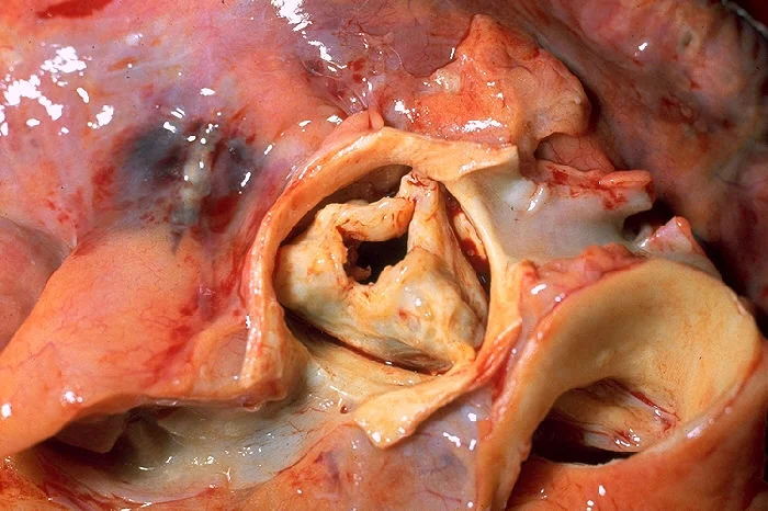
- Degenerative calcification - the commonest cause over 65. An active inflammatory and calcific process sharing risk factors with atherosclerosis, not simple wear and tear.
- Bicuspid aortic valve - the commonest cause under 65, and the commonest congenital cardiac abnormality, present in 1-2% of the population. Two leaflets instead of three means abnormal flow and calcification 10-20 years earlier than a normal valve.
- Rheumatic heart disease - commonest worldwide; usually with coexisting mitral disease
- Supravalvular or subvalvular obstruction - including hypertrophic obstructive cardiomyopathy, which mimics aortic stenosis
Pathophysiology
Obstruction forces the left ventricle to generate a much higher pressure to eject blood. It compensates with concentric hypertrophy - a thicker wall around a normal or small cavity. This maintains output for years but has three consequences that map exactly onto the symptoms:
- The thickened muscle needs more oxygen, while the raised intramural pressure impairs coronary perfusion during diastole - producing angina even with normal coronary arteries
- Cardiac output becomes fixed, so it cannot rise on exertion when peripheral vessels dilate - producing exertional syncope
- The stiff ventricle fills poorly and eventually fails - producing dyspnoea and heart failure
Aortic stenosis: clinical features
Aortic stenosis is asymptomatic for decades. The onset of symptoms is the single most important event in its natural history, because it marks a sharp change in prognosis.
Examination
- Pulse: slow-rising and low volume - pulsus parvus et tardus. Feel it at the carotid, where the character is clearest.
- Blood pressure: narrow pulse pressure
- Apex beat: heaving and not displaced, because the ventricle is thick rather than dilated
- Thrill in the aortic area, and a palpable carotid thrill (shudder)
- Murmur: ejection systolic, crescendo-decrescendo, loudest in the 2nd right intercostal space, radiating to the carotids, louder on expiration and leaning forward
- Second heart sound: soft or absent as the calcified valve stops closing audibly - a useful marker of severity
- Fourth heart sound from forceful atrial contraction into a stiff ventricle
- Signs of heart failure in decompensated disease
Aortic regurgitation
Aortic regurgitation results either from disease of the valve leaflets or from dilatation of the aortic root, which pulls the leaflets apart so they no longer meet.
| Acute | Chronic |
|---|---|
| Infective endocarditis | Bicuspid aortic valve |
| Aortic dissection | Rheumatic heart disease |
| Chest trauma | Aortic root dilatation - Marfan syndrome, Ehlers-Danlos syndrome |
| Failure of a prosthetic valve | Ankylosing spondylitis and other seronegative arthritides |
| Hypertension; previously, tertiary syphilis |
Why acute and chronic behave so differently
In chronic regurgitation the ventricle has years to adapt, dilating progressively to accommodate the extra volume while keeping filling pressures near normal. Patients stay well for a very long time, then decompensate.
In acute regurgitation there is no time to adapt. A normal-sized, non-compliant ventricle suddenly receives a large regurgitant volume, so left ventricular end-diastolic pressure rises abruptly and is transmitted to the lungs. The patient presents with sudden severe pulmonary oedema and cardiogenic shock, and characteristically the murmur is short and soft - the very high ventricular pressure quickly equalises with aortic pressure, so there is little to hear. It is easy to underestimate, and it is a surgical emergency.
Examination in chronic aortic regurgitation
- Pulse: collapsing or water-hammer - felt best with the arm raised
- Blood pressure: wide pulse pressure, with a low diastolic
- Apex beat: displaced, diffuse and hyperdynamic (volume-loaded)
- Murmur: early diastolic, high-pitched and blowing, at the left sternal edge, loudest with the patient sitting forward in held expiration
- Austin Flint murmur - a mid-diastolic rumble at the apex, caused by the regurgitant jet striking the anterior mitral leaflet
- An ejection systolic flow murmur may coexist, from the increased stroke volume crossing the valve
Investigations
Echocardiography is the definitive investigation for both lesions. It confirms the diagnosis, grades severity, and measures the ventricular consequences that determine the timing of surgery.
| Measure | Severe |
|---|---|
| Aortic valve area | Below 1.0 cm² |
| Mean pressure gradient | 40 mmHg or above |
| Peak aortic jet velocity | 4.0 m/s or above |
Note that gradients depend on flow: a patient with a poor ejection fraction may have severe stenosis with a low gradient, because the ventricle cannot generate one. This 'low-flow, low-gradient' pattern is easily missed and may need dobutamine stress echocardiography to clarify.
- ECG - left ventricular hypertrophy with strain in stenosis; left axis deviation; conduction disease, since the calcified valve sits next to the conducting system
- Chest X-ray - a relatively normal heart size in stenosis until late (the ventricle thickens rather than dilates), a calcified valve, post-stenotic aortic dilatation; cardiomegaly in chronic regurgitation
- Bloods - FBC, U&Es, NT-proBNP, and blood cultures if endocarditis is possible
- Cardiac CT - valve calcium scoring, and essential planning for TAVI including access vessels
- Cardiac MRI - quantifies regurgitant fraction and assesses the aortic root
- Coronary angiography - before intervention, as coexisting coronary disease is common and can be treated at the same time
- Exercise testing - used cautiously in apparently asymptomatic severe stenosis to unmask symptoms. It is contraindicated in symptomatic severe aortic stenosis.
Management
Aortic stenosis
No medical therapy alters the course of aortic stenosis. Statins do not slow calcification. Management consists of surveillance, careful treatment of comorbidity, and valve replacement at the right moment.
Indications for intervention:
- Symptomatic severe aortic stenosis - the principal indication
- Asymptomatic severe stenosis with a left ventricular ejection fraction below 50%
- Asymptomatic severe stenosis with an abnormal exercise test - symptoms or a fall in blood pressure on exertion
- Severe stenosis in a patient undergoing cardiac surgery for another reason
| Option | Notes |
|---|---|
| Surgical aortic valve replacement (SAVR) | Open surgery with a mechanical or bioprosthetic valve. Preferred in younger, lower-risk patients, and where the aortic root also needs surgery. |
| Transcatheter aortic valve implantation (TAVI) | Delivered via the femoral artery, usually without general anaesthesia. Originally reserved for those unsuitable for surgery; now used across an increasingly wide range of surgical risk, particularly in older patients. |
| Balloon aortic valvuloplasty | Temporary relief only, with rapid restenosis. Used as a bridge in unstable patients or before non-cardiac surgery. |
TAVI carries a higher rate of conduction disturbance requiring a permanent pacemaker and of paravalvular leak; surgery carries the risks of sternotomy and bypass. The choice is made by a heart team weighing age, frailty, anatomy and comorbidity.
Aortic regurgitation
Chronic: treat hypertension, which reduces the regurgitant volume - vasodilators such as ACE inhibitors, ARBs or dihydropyridine calcium channel blockers are used here, in contrast to stenosis. Follow with serial echocardiography. Where the aortic root is dilated, particularly in Marfan syndrome, a beta-blocker or ARB slows expansion and the threshold for surgery is lower.
Indications for surgery in chronic regurgitation: symptoms; or, in an asymptomatic patient, a left ventricular ejection fraction of 50% or below, or progressive left ventricular dilatation. Root replacement is added where the aorta is aneurysmal.
Complications
Both lesions converge on left ventricular failure if untreated, but each has characteristic additional complications.
- Sudden cardiac death - notably in symptomatic severe aortic stenosis
- Heart failure - the common endpoint of both lesions
- Atrial fibrillation and other arrhythmias
- Conduction disease and complete heart block - from calcium extending into the conducting system, and after TAVI or surgery
- Infective endocarditis - the abnormal valve is a substrate, and bicuspid valves are at particular risk
- Aortic dissection and aneurysm - especially with a bicuspid valve or connective tissue disease
- Acquired von Willebrand syndrome with angiodysplasia - Heyde syndrome, causing gastrointestinal bleeding in severe aortic stenosis, and it resolves after valve replacement
- Prosthetic valve complications - thrombosis, degeneration, paravalvular leak and haemolysis
Red flags
Prognosis
Asymptomatic aortic stenosis carries a low annual risk of sudden death, and patients can be safely followed with serial echocardiography. Once symptoms develop the picture changes abruptly - untreated symptomatic severe stenosis has a worse prognosis than most malignancies, with average survival of two to five years depending on the symptom.
Valve replacement transforms this. Operative mortality for isolated aortic valve replacement is low in suitable candidates, and long-term survival after successful replacement approaches that of the general population of the same age. The introduction of TAVI has extended that benefit to patients who would previously have been managed palliatively.
Chronic aortic regurgitation has a long compensated phase, and asymptomatic patients with normal ventricular function do well for many years. The determinant of outcome is operating before irreversible ventricular dilatation and dysfunction develop - which is precisely why asymptomatic patients are monitored with echocardiography rather than simply reassured and discharged.
References
- Vahanian A, Beyersdorf F, Praz F et al. ESC/EACTS Guidelines for the management of valvular heart disease. European Heart Journal. 2021. Available here
- NICE NG208. Heart valve disease presenting in adults: investigation and management. 2021. Available here
- Ross J, Braunwald E. Aortic stenosis. Circulation. 1968. Available here
- NICE IPG586. Transcatheter aortic valve implantation for aortic stenosis. 2017. Available here
- NICE Clinical Knowledge Summaries. Heart murmurs. Available here
- Otto CM, Nishimura RA, Bonow RO et al. ACC/AHA Guideline for the management of patients with valvular heart disease. Circulation. 2021. Available here
- BNF. Glyceryl trinitrate - cautions and contraindications. Available here
This article is written for revision and education. It is not clinical guidance and must not be used to make decisions about the care of a patient. Always check current NICE guidance and local protocols.