
Valvular Heart Disease: An Approach to Murmurs
Key points
- Valvular heart disease: narrowing (stenosis) or leaking (regurgitation) of one or more of the four heart valves.
- Commonest causes: degenerative calcification in the UK; rheumatic heart disease remains the commonest cause worldwide.
- Timing first: systolic murmurs are aortic or pulmonary stenosis, or mitral or tricuspid regurgitation. Diastolic murmurs are the reverse pairing.
- RILE: Right-sided murmurs are louder on Inspiration, Left-sided on Expiration.
- A diastolic murmur is never innocent: it always warrants echocardiography.
- Investigation: echocardiography is the definitive test, and it grades severity as well as identifying the lesion.
- Antibiotic prophylaxis: NICE does not recommend routine antibiotic prophylaxis against infective endocarditis for dental or other procedures.
- Mechanical valves: require lifelong warfarin. DOACs are contraindicated.
Introduction
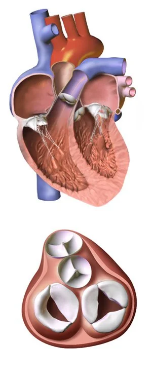
The four cardiac valves keep blood moving in one direction. A valve can fail in two ways: it can fail to open properly (stenosis), obstructing forward flow and imposing a pressure load on the chamber behind it; or it can fail to close properly (regurgitation), allowing backward flow and imposing a volume load.
That distinction explains nearly everything downstream. Pressure overload produces concentric hypertrophy - a thick-walled, non-dilated chamber, as in aortic stenosis. Volume overload produces eccentric hypertrophy - a dilated chamber, as in aortic or mitral regurgitation. The pattern determines the symptoms, the examination findings and the timing of surgery.
Valvular disease affects over 10% of people above 75 in the UK and is becoming more common as the population ages. It is also one of the highest-yield examination topics, because the diagnosis can genuinely be made at the bedside.
Aetiology
| Cause | Typical picture |
|---|---|
| Degenerative calcification | The commonest cause in the UK. Age-related, sharing risk factors with atherosclerosis. Mainly aortic stenosis and mitral annular calcification. |
| Rheumatic heart disease | The commonest cause worldwide. Follows group A streptococcal pharyngitis. Affects the mitral valve most, then aortic. |
| Congenital | Bicuspid aortic valve is the commonest congenital cardiac abnormality, present in 1-2% of people, and it calcifies decades earlier than a tricuspid valve. |
| Infective endocarditis | Destroys leaflets and causes acute regurgitation. Suspect in any new murmur with fever. |
| Connective tissue disease | Marfan and Ehlers-Danlos syndromes cause aortic root dilatation and mitral valve prolapse. |
| Functional or secondary | The valve itself is normal but the annulus is stretched by ventricular dilatation - common in heart failure, causing functional mitral and tricuspid regurgitation. |
| Other | Radiotherapy, carcinoid syndrome (right-sided), systemic lupus erythematosus (Libman-Sacks), and previously syphilis (aortic regurgitation). |
Clinical features
Valvular disease is often silent for years, because the ventricle compensates. Symptoms appear when compensation fails, and their onset is a critical point in the natural history - in aortic stenosis it is the trigger for surgery.
- Breathlessness on exertion, then orthopnoea and paroxysmal nocturnal dyspnoea - the commonest symptom of most lesions
- Chest pain - typically in aortic stenosis, from increased myocardial demand with reduced coronary perfusion
- Syncope or pre-syncope on exertion - a red flag, classically in severe aortic stenosis
- Palpitations - atrial fibrillation is common, particularly in mitral disease where the left atrium dilates
- Fatigue and reduced exercise tolerance
- Ankle swelling and abdominal distension - right-sided lesions or advanced left-sided disease
- Haemoptysis - classically in mitral stenosis, from raised pulmonary venous pressure
Ask specifically about rheumatic fever or frequent sore throats in childhood, intravenous drug use, previous valve surgery, dental care, and a family history of sudden death or connective tissue disease.
Examining a murmur
A systematic approach makes murmurs far more tractable than they first appear. Work through: timing, site, radiation, character, intensity, and what changes it.
Where to listen
| Valve | Where | Radiates to |
|---|---|---|
| Aortic | 2nd intercostal space, right sternal edge | Carotids |
| Pulmonary | 2nd intercostal space, left sternal edge | - |
| Tricuspid | 4th-5th intercostal space, left sternal edge | - |
| Mitral | 5th intercostal space, mid-clavicular line (the apex) | Axilla |
Timing narrows it to two
| Systolic | Diastolic |
|---|---|
| Aortic stenosis | Aortic regurgitation |
| Pulmonary stenosis | Pulmonary regurgitation |
| Mitral regurgitation | Mitral stenosis |
| Tricuspid regurgitation | Tricuspid stenosis |
| Ventricular septal defect; hypertrophic obstructive cardiomyopathy | - |
The logic is simple: in systole the ventricles contract, so blood should be leaving through the aortic and pulmonary valves and the mitral and tricuspid valves should be shut. A systolic murmur therefore means either an obstructed outflow valve or a leaking inflow valve. Diastole reverses the pairing.
Grading intensity
The Levine scale grades murmurs 1 to 6. Grade 1 is barely audible; grade 2 is quiet but clearly heard; grade 3 is moderately loud with no thrill; grade 4 is the first with a palpable thrill; grade 5 is audible with the stethoscope barely touching the chest; grade 6 is audible with it lifted off. The presence of a thrill is the reproducible landmark - and intensity correlates poorly with severity, since a critically stenotic valve may generate a quiet murmur because so little blood is crossing it.
Investigations
- Transthoracic echocardiography - the definitive investigation. It identifies which valve, whether stenosis or regurgitation, grades severity, and measures the consequences: chamber sizes, ejection fraction and pulmonary pressures.
- Transoesophageal echocardiography (TOE) - better views of the mitral valve, prosthetic valves and vegetations; used where transthoracic images are inadequate and in suspected endocarditis
- ECG - left ventricular hypertrophy in aortic stenosis; left atrial enlargement (P mitrale) and atrial fibrillation in mitral disease; right ventricular hypertrophy in pulmonary and tricuspid disease
- Chest X-ray - cardiomegaly, pulmonary congestion, a calcified valve, and left atrial enlargement
- Bloods - FBC and inflammatory markers, blood cultures if endocarditis is suspected, U&Es, and NT-proBNP
- Cardiac CT or MRI - valve calcium scoring, aortic root assessment, and quantifying regurgitation
- Coronary angiography - performed before valve surgery in anyone with risk factors, since coexisting coronary disease is common and can be grafted at the same operation
- Exercise testing - carefully used in apparently asymptomatic severe aortic stenosis to unmask symptoms
Management
No drug reverses valvular disease. Medical therapy manages the consequences - heart failure, atrial fibrillation, hypertension - while the definitive question is whether and when to intervene.
Surveillance
Asymptomatic patients with mild or moderate disease are followed with serial echocardiography, the interval depending on the lesion and its severity - typically every 3-5 years for mild disease and annually for severe. The purpose is to catch the transition to symptoms or to ventricular decompensation before irreversible damage occurs.
Deciding to intervene
Two triggers dominate across all lesions: the onset of symptoms, and evidence that the ventricle is failing - a falling ejection fraction or progressive dilatation - even in a patient who feels well. Decisions are made by a multidisciplinary heart team.
| Procedure | Used for |
|---|---|
| Surgical valve replacement | The established option for most lesions; requires sternotomy and bypass |
| Valve repair | Preferred to replacement where feasible, particularly for the mitral valve - it preserves the native valve and avoids anticoagulation |
| Transcatheter aortic valve implantation (TAVI) | Aortic stenosis, delivered via the femoral artery. Originally for those unsuitable for surgery, now used across a widening range of surgical risk. |
| Transcatheter edge-to-edge repair | Selected patients with severe mitral regurgitation who are poor surgical candidates |
| Balloon valvuloplasty | Mitral stenosis with suitable valve anatomy; and as a temporising bridge in aortic stenosis |
Choosing a prosthetic valve
| Mechanical | Bioprosthetic | |
|---|---|---|
| Durability | Lasts a lifetime | Structural degeneration over 10-20 years |
| Anticoagulation | Lifelong warfarin, with a target INR set by valve type and position | Usually only short-term antiplatelet or anticoagulation |
| Best suited to | Younger patients, where durability matters most | Older patients, and anyone in whom anticoagulation is hazardous or unwanted |
| Sound | An audible click | Silent |
Complications
- Heart failure - the common endpoint of every untreated significant lesion
- Atrial fibrillation - particularly with mitral disease and left atrial dilatation, bringing its own stroke risk
- Infective endocarditis - abnormal and prosthetic valves are both at increased risk
- Pulmonary hypertension and right heart failure - from chronically raised left-sided pressures
- Systemic embolism - from left atrial thrombus in mitral stenosis with atrial fibrillation, or from valve vegetations
- Sudden cardiac death - notably in severe aortic stenosis, especially once symptomatic
- Prosthetic valve complications - thrombosis, structural degeneration, paravalvular leak, haemolysis and endocarditis
Red flags
Prognosis
Prognosis varies enormously with the lesion, its severity and whether it has been corrected. Mild asymptomatic disease may never progress to needing intervention; severe symptomatic aortic stenosis left untreated has an outlook worse than many cancers.
The general rule is that valve replacement performed before the ventricle decompensates restores a near-normal life expectancy, whereas surgery performed after irreversible ventricular damage does not. This is why asymptomatic patients are followed with serial imaging rather than discharged, and why a falling ejection fraction is an indication to operate even in someone who feels well.
The arrival of transcatheter techniques has substantially changed the outlook for older and frailer patients, many of whom were previously offered nothing because open surgery was too risky.
References
- Vahanian A, Beyersdorf F, Praz F et al. ESC/EACTS Guidelines for the management of valvular heart disease. European Heart Journal. 2021. Available here
- NICE CG64. Prophylaxis against infective endocarditis. 2008, updated 2016. Available here
- Eikelboom JW, Connolly SJ, Brueckmann M et al. Dabigatran versus warfarin in patients with mechanical heart valves (RE-ALIGN). New England Journal of Medicine. 2013. Available here
- NICE NG208. Heart valve disease presenting in adults: investigation and management. 2021. Available here
- NICE Clinical Knowledge Summaries. Heart murmurs. Available here
- World Health Organization. Rheumatic heart disease fact sheet. Available here
- BNF. Warfarin sodium - indications and target INR. Available here
This article is written for revision and education. It is not clinical guidance and must not be used to make decisions about the care of a patient. Always check current NICE guidance and local protocols.