
Hypertrophic Obstructive Cardiomyopathy: Diagnosis and Management
Key points
- HOCM: an autosomal dominant sarcomeric disorder causing left ventricular hypertrophy that is not explained by loading conditions.
- Frequency: the commonest inherited cardiac condition, affecting around 1 in 500, and the leading cause of sudden cardiac death in young athletes.
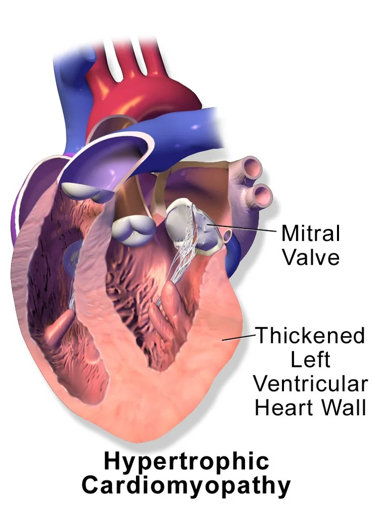
- Obstruction: asymmetric septal hypertrophy plus systolic anterior motion of the mitral valve narrows the outflow tract during systole.
- The murmur: ejection systolic at the left sternal edge that does NOT radiate to the carotids, and gets louder on Valsalva or standing.
- Red flag symptom: exertional syncope. It suggests obstruction or arrhythmia and demands urgent assessment.
- Avoid: vasodilators, nitrates, ACE inhibitors and digoxin - anything that reduces preload or afterload worsens the obstruction.
- First-line drug: a beta-blocker; verapamil second line; disopyramide added for refractory symptoms.
- Family screening: offer genetic counselling and cardiac screening to all first-degree relatives.
Introduction
Hypertrophic cardiomyopathy is left ventricular hypertrophy that cannot be explained by the loading conditions of the heart - so it is not the hypertrophy of hypertension or aortic stenosis. It is defined by a wall thickness of 15 mm or more in an adult, or 13 mm or more in a first-degree relative of an affected patient.1
It is the commonest inherited cardiac condition, affecting around 1 in 500 people, and the commonest cause of sudden cardiac death in young people and athletes. Most patients, however, have a normal life expectancy - and the central clinical task is identifying the minority at high risk.
The word obstructive refers to dynamic obstruction of the left ventricular outflow tract, present at rest in around a third of patients and provoked by exertion in another third. The remainder have non-obstructive disease. Because the obstruction is dynamic rather than fixed, it varies from beat to beat with loading conditions - which explains almost everything distinctive about the condition.
Aetiology
HOCM is a disease of the sarcomere, the contractile unit of the myocyte. It is inherited in an autosomal dominant pattern with variable penetrance and expression - so a mutation carrier may be severely affected, mildly affected, or apparently normal.
- MYH7 - beta-myosin heavy chain
- MYBPC3 - myosin-binding protein C
- Together these account for the majority of identified mutations; over 1,500 variants have been described across at least 11 sarcomeric genes
- Around 40-60% of patients have an identifiable sarcomeric mutation; the remainder have no genetic diagnosis
Pathophysiology
Four consequences follow from the abnormal muscle, and between them they explain every symptom:
- Outflow tract obstruction - asymmetric hypertrophy of the interventricular septum narrows the outflow tract. Rapid flow past the mitral valve drags the anterior leaflet towards the septum (systolic anterior motion, SAM), worsening the obstruction and simultaneously causing mitral regurgitation.
- Diastolic dysfunction - the thickened, stiff ventricle fills poorly, raising filling pressures and causing breathlessness.
- Myocardial ischaemia - the hypertrophied muscle outgrows its blood supply, and small intramural coronary arteries are abnormal. Angina occurs with normal epicardial coronary arteries.
- Myocyte disarray and fibrosis - the myocytes are arranged chaotically rather than in parallel, creating a substrate for ventricular arrhythmia and sudden death.
Risk factors
The dominant risk factor is genetic: a first-degree relative with HOCM confers a 50% chance of inheriting the mutation. There are no modifiable risk factors for developing the condition.
What is modifiable is the risk of an adverse event in someone who has it:
- Competitive or intense sport - the classic trigger for sudden cardiac death
- Dehydration and hypovolaemia - reduced preload increases obstruction
- Vasodilating drugs - nitrates, ACE inhibitors, dihydropyridine calcium channel blockers
- Excessive diuresis
- Alcohol and vasodilating environments such as hot baths
- Atrial fibrillation - loss of the atrial contribution to filling is poorly tolerated by a stiff ventricle
Clinical features
Many patients are asymptomatic and are identified through family screening, an incidental murmur, or an abnormal ECG. Tragically, in some the first manifestation is sudden death.
- Exertional breathlessness - the commonest symptom, from diastolic dysfunction and obstruction
- Chest pain - typically exertional angina despite normal coronary arteries
- Palpitations - atrial fibrillation, or ventricular arrhythmia
- Syncope and pre-syncope - especially on exertion; a red flag suggesting severe obstruction or ventricular arrhythmia
- Sudden cardiac death - often during or just after exertion, and sometimes the presenting event
- Symptoms characteristically worsen with dehydration, standing suddenly, after a large meal, after alcohol, and following vasodilators
Take a careful family history across three generations, asking specifically about sudden death, unexplained death in infancy, drowning, single-vehicle road accidents, epilepsy diagnoses and pacemaker or defibrillator implantation in relatives - all of which may represent undiagnosed inherited cardiac disease.
Clinical examination
Examination may be entirely normal, particularly in non-obstructive disease. Where obstruction is present, the findings are distinctive:
- Pulse: jerky with a sharp upstroke - the ventricle empties rapidly before the obstruction develops mid-systole. Sometimes bisferiens (two peaks).
- Apex beat: forceful and double or triple impulse, from a palpable fourth heart sound as the atrium contracts against a stiff ventricle
- Murmur: ejection systolic, harsh, loudest at the lower left sternal edge and apex, and characteristically not radiating to the carotids
- A pansystolic murmur at the apex radiating to the axilla may coexist, from the mitral regurgitation caused by systolic anterior motion
- Fourth heart sound (S4)
- The JVP may show a prominent a wave
Differential diagnosis
- Aortic stenosis - the key clinical differential; separated by the manoeuvres above and by echocardiography
- Hypertensive left ventricular hypertrophy - symmetrical, usually under 15 mm, with a clear history of hypertension
- Athlete's heart - physiological, symmetrical, with a dilated cavity and normal diastolic function
- Cardiac amyloidosis - low ECG voltages with thick walls on echo is the tell-tale mismatch
- Fabry disease - X-linked, with extracardiac features and a specific treatment
- Aortic subvalvular membrane - fixed subaortic obstruction that mimics HOCM
- Coronary artery disease - for the chest pain
- Vasovagal syncope and epilepsy - for the syncope; exertional syncope in a young person should never be attributed to either without cardiac assessment
Investigations
ECG
Abnormal in more than 90% of patients, and often the finding that prompts the diagnosis. It is rarely normal, which makes it a useful screening test in relatives.
- Left ventricular hypertrophy by voltage criteria
- Deep, narrow Q waves in the inferior and lateral leads - so-called 'dagger' Q waves from septal hypertrophy, and easily mistaken for previous infarction
- T wave inversion, particularly inferolaterally
- Giant T wave inversion in the precordial leads - characteristic of apical hypertrophic cardiomyopathy
- Left atrial enlargement; atrial fibrillation; occasionally a short PR interval with a delta wave
Echocardiography
The primary diagnostic test. Look for:
- Asymmetric septal hypertrophy with a wall thickness of 15 mm or more (13 mm in a relative)
- Systolic anterior motion (SAM) of the anterior mitral leaflet
- Left ventricular outflow tract gradient - a resting or provoked gradient of 30 mmHg or more defines obstruction; 50 mmHg or more is the threshold for considering septal reduction therapy
- Mitral regurgitation from SAM, with a posteriorly directed jet
- A small, hyperdynamic left ventricular cavity with impaired diastolic filling
If no gradient is present at rest, provocation with Valsalva, standing or exercise is needed - resting echocardiography alone misses a third of obstructive cases.
Further investigations
- Cardiac MRI - superior for apical and lateral wall hypertrophy that echo can miss, and quantifies late gadolinium enhancement (fibrosis), which is an emerging marker of arrhythmic risk
- Ambulatory ECG monitoring (48 hours or longer) - to detect non-sustained ventricular tachycardia, a key component of risk stratification, and atrial fibrillation
- Exercise testing - assesses functional capacity, provokes latent obstruction, and looks for an abnormal blood pressure response (a failure to rise, or a fall) which carries prognostic weight
- Genetic testing - identifies a causative mutation in around half, and once found allows cascade screening of relatives with a definitive yes or no
- Bloods - NT-proBNP, and investigations for phenocopies where suspected: serum free light chains and bone scintigraphy for amyloidosis, alpha-galactosidase A for Fabry disease
Management
Management has three separate goals that should be considered in turn: relieve symptoms, prevent sudden death, and screen the family. They are largely independent - a patient with mild symptoms may still be at high arrhythmic risk.
Lifestyle and things to avoid
Drug treatment of symptoms
- Beta-blockers - first line. They slow the heart, lengthening diastolic filling time, and reduce contractility and therefore the gradient.
- Verapamil - second line where beta-blockers are not tolerated. Avoid in severe obstruction or resting gradients above 100 mmHg, and never combine it with a beta-blocker.
- Disopyramide - added for symptoms refractory to a beta-blocker; strongly negatively inotropic, though its antimuscarinic effects limit tolerability
- Cardiac myosin inhibitors (mavacamten) - a newer class that targets the underlying excessive actin-myosin cross-bridging, reducing gradients and improving symptoms; used in specialist centres with echocardiographic monitoring
- Cautious diuretics for congestion in non-obstructive disease
Septal reduction therapy
For patients with severe symptoms and a gradient of 50 mmHg or more despite optimal drugs:
- Surgical septal myectomy - excision of the hypertrophied septum. Excellent long-term results in experienced centres, and preferred in younger patients or where the mitral valve also needs surgery.
- Alcohol septal ablation - injection of alcohol into a septal perforator artery to infarct the septum. Less invasive, suits older patients or those unfit for surgery. Carries a significant risk of complete heart block requiring a permanent pacemaker.
Preventing sudden cardiac death
An implantable cardioverter defibrillator (ICD) is the only effective measure. Secondary prevention - after a cardiac arrest or sustained ventricular tachycardia - is straightforward. Primary prevention requires formal risk estimation, using the HCM Risk-SCD score, which combines:1
- Age
- Maximal left ventricular wall thickness
- Left atrial diameter
- Maximal left ventricular outflow tract gradient
- Family history of sudden cardiac death
- Non-sustained ventricular tachycardia on ambulatory monitoring
- Unexplained syncope
Atrial fibrillation
Family screening
Offer genetic counselling and cardiac assessment - ECG and echocardiography - to all first-degree relatives. Where a causative mutation is found in the index patient, cascade genetic testing gives relatives a definitive answer and allows those without the mutation to be discharged. Where no mutation is identified, relatives need periodic clinical screening through adolescence and into adulthood, since the phenotype may appear later.
Complications
- Sudden cardiac death - from ventricular arrhythmia; the most feared complication, and often the presenting event in young people
- Atrial fibrillation - affects around a quarter of patients, poorly tolerated, and carries a high stroke risk
- Stroke and systemic embolism
- Progressive heart failure - initially with preserved ejection fraction from diastolic dysfunction
- End-stage 'burnt-out' disease - a minority progress to a dilated, thin-walled, poorly contracting ventricle, which may require transplantation
- Infective endocarditis - uncommon, and related to the mitral regurgitation and turbulent outflow
- Complete heart block - a recognised consequence of alcohol septal ablation
Red flags
Prognosis
The prognosis is far better than its reputation suggests. Contemporary cohorts report an annual mortality of around 0.5-1%, and the majority of patients have a normal life expectancy with no significant limitation.2 The historical figures came from specialist referral centres and were heavily skewed towards the most severe cases.
Outcomes have improved substantially with ICDs for those at high arrhythmic risk, effective septal reduction for obstruction, anticoagulation for atrial fibrillation, and systematic family screening that identifies affected relatives before they become symptomatic.
Adverse features are a family history of sudden death, unexplained syncope, non-sustained ventricular tachycardia, extreme wall thickness, extensive fibrosis on cardiac MRI, and progression to end-stage disease. The practical message for a student is that the diagnosis is not a death sentence - but exertional syncope, a family history of sudden death, or an abnormal ECG in a young athlete all demand assessment rather than reassurance.
References
- Arbelo E, Protonotarios A, Gimeno JR et al. ESC Guidelines for the management of cardiomyopathies. European Heart Journal. 2023. Available here
- Maron BJ, Rowin EJ, Casey SA et al. Hypertrophic cardiomyopathy in adulthood associated with low cardiovascular mortality. Journal of the American College of Cardiology. 2015. Available here
- O'Mahony C, Jichi F, Pavlou M et al. A novel clinical risk prediction model for sudden cardiac death in hypertrophic cardiomyopathy (HCM Risk-SCD). European Heart Journal. 2014. Available here
- NICE Clinical Knowledge Summaries. Cardiomyopathy. Available here
- NICE IPG317. Percutaneous alcohol septal ablation for hypertrophic obstructive cardiomyopathy. 2009. Available here
- Cardiomyopathy UK. Hypertrophic cardiomyopathy information. Available here
- BNF. Bisoprolol fumarate - indications and dosing. Available here
This article is written for revision and education. It is not clinical guidance and must not be used to make decisions about the care of a patient. Always check current NICE guidance and local protocols.