
Polymyositis and Dermatomyositis
Key points
- Polymyositis and dermatomyositis: idiopathic inflammatory myopathies causing symmetrical, insidious-onset proximal muscle weakness - difficulty rising from a chair, climbing stairs, or raising the arms above the head.
- Dermatomyositis-specific signs: heliotrope rash (violaceous eyelid discolouration), Gottron's papules (scaly papules over the knuckles - near pathognomonic), and the shawl sign (photosensitive erythema across the shoulders and upper back).
- Amyopathic dermatomyositis: the characteristic skin changes can occur without significant muscle weakness - do not exclude the diagnosis because strength is preserved.
- Malignancy association: dermatomyositis, especially in older patients and with anti-TIF1-gamma antibody, carries a substantially increased risk of an underlying malignancy - screen at diagnosis.
- Key investigation: creatine kinase is markedly elevated, often more than 10 times the upper limit of normal, and tracks disease activity.
- Gold standard: muscle biopsy - perifascicular atrophy in dermatomyositis versus endomysial inflammatory infiltrate in polymyositis.
- Antisynthetase syndrome: myositis, interstitial lung disease, arthritis, mechanic's hands, Raynaud phenomenon and fever, associated with anti-Jo-1 and other anti-synthetase antibodies.
- First-line treatment: high-dose corticosteroids, with a steroid-sparing immunosuppressant started early given the burden of long-term steroid use.
Introduction
Polymyositis (PM) and dermatomyositis (DM) are the two principal idiopathic inflammatory myopathies: autoimmune diseases in which the immune system attacks skeletal muscle, producing symmetrical proximal muscle weakness. Dermatomyositis is distinguished by a set of characteristic skin findings that can precede, accompany, or - in amyopathic disease - occur without significant muscle involvement.1
They sit within a broader family of inflammatory myopathies that also includes inclusion body myositis (typically older patients, distal as well as proximal weakness, poor response to immunosuppression) and immune-mediated necrotising myopathy (often anti-SRP or statin-associated, severe weakness with very high CK), but PM and DM are the two that dominate exam content.
The two features that make this topic reliably testable are the characteristic dermatomyositis rashes, which are genuinely pattern-recognition medicine, and the paraneoplastic association of dermatomyositis, which changes what you actually do at diagnosis.
Aetiology
Both conditions are autoimmune, but the underlying immunopathology differs: dermatomyositis is primarily a humoral, complement-mediated microangiopathy affecting the perimysium and perifascicular muscle fibres, while polymyositis involves direct cytotoxic T-cell attack on muscle fibres (endomysial inflammation). This distinction, visible on biopsy, is why the two conditions - though clinically similar - are considered pathologically distinct.
Peak onset is in adulthood (40s-60s), though juvenile dermatomyositis occurs in children and carries its own distinct pattern, including a higher rate of calcinosis and a much lower malignancy association.
Clinical features
Muscle weakness
- Symmetrical, proximal muscle weakness, developing insidiously over weeks to months
- Difficulty rising from a low chair, climbing stairs, or raising the arms above the head (to brush hair, for example)
- Myalgia in some patients, though weakness rather than pain is the dominant symptom
- Neck flexor weakness and, in more severe disease, dysphagia from pharyngeal muscle involvement
- Preserved reflexes and sensation - this is a myopathy, not a neuropathy, which helps distinguish it from Guillain-Barre syndrome or a peripheral neuropathic cause of weakness
Dermatomyositis-specific skin signs
- Heliotrope rash - a violaceous discolouration of the upper eyelids, often with periorbital oedema
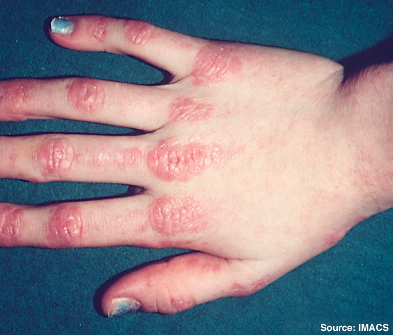
- Gottron's papules - scaly, erythematous or violaceous papules over the extensor surfaces of the metacarpophalangeal and interphalangeal joints - near pathognomonic for dermatomyositis
- Gottron's sign - the same erythema without discrete papules, over the elbows, knees and knuckles
- Shawl sign - photosensitive erythema across the back, shoulders and upper chest in a shawl-like distribution
- Mechanic's hands - cracked, fissured, hyperkeratotic skin along the lateral fingers, particularly associated with antisynthetase syndrome
- Periungual (nailfold) erythema and capillary changes
- Calcinosis cutis - subcutaneous calcium deposits, especially common in juvenile dermatomyositis
Extramuscular and systemic features
- Interstitial lung disease - particularly associated with antisynthetase syndrome and anti-MDA5 antibody, and can be rapidly progressive
- Cardiac involvement - myocarditis and conduction abnormalities
- Arthralgia/arthritis
- Raynaud phenomenon
- Fever and constitutional symptoms, particularly in antisynthetase syndrome
Antisynthetase syndrome
Antisynthetase syndrome is a recognisable overlap phenotype, driven by antibodies against aminoacyl-tRNA synthetases (most often anti-Jo-1), combining myositis, interstitial lung disease, inflammatory arthritis, mechanic's hands, Raynaud phenomenon and fever. Interstitial lung disease is often the feature that dominates the clinical course and drives long-term morbidity in this subgroup.
Malignancy association
Dermatomyositis carries a well-established association with underlying malignancy, particularly in patients over 40-50, with the risk highest in the year before and after diagnosis. Ovarian, lung, pancreatic, gastric, colorectal cancer and lymphoma are the most commonly reported associations.2
Differential diagnosis
- Statin-induced myopathy - myalgia and mild CK elevation are common with statins; a markedly raised CK with true weakness should prompt consideration of immune-mediated necrotising myopathy (which can be triggered by statins and persists after stopping them)
- Inclusion body myositis - older patients, distal as well as proximal weakness (notably finger flexors and quadriceps), asymmetrical, poor steroid response
- Polymyalgia rheumatica - pain and stiffness rather than true weakness, normal CK
- Hypothyroidism - can cause proximal weakness and a raised CK, easily distinguished with TFTs
- Guillain-Barre syndrome and other neuropathies - weakness with reduced or absent reflexes, unlike the preserved reflexes of myopathy
- Muscular dystrophy - usually presents earlier in life with a clear inheritance pattern
Investigations
- Creatine kinase (CK) - typically markedly elevated, often more than 10 times the upper limit of normal, and used to track disease activity and treatment response
- Aldolase, LDH, AST/ALT - also released from damaged muscle, and can be mistaken for liver disease if CK is not checked
- Myositis-specific and myositis-associated antibody panel - anti-Jo-1 (antisynthetase syndrome), anti-Mi-2 (classic DM, generally good prognosis), anti-TIF1-gamma (malignancy association), anti-SRP (severe necrotising myopathy), anti-MDA5 (DM with a risk of rapidly progressive, life-threatening ILD, and often amyopathic skin-predominant disease)
- EMG - a myopathic pattern of short-duration, low-amplitude, polyphasic motor unit potentials, with spontaneous fibrillations
- MRI of muscle - shows oedema and inflammation, useful both to support the diagnosis and to identify a good biopsy target
- Muscle biopsy - the gold standard, showing perifascicular atrophy with a perivascular/perimysial inflammatory infiltrate in dermatomyositis, versus endomysial inflammatory infiltrate surrounding and invading individual muscle fibres in polymyositis
- High-resolution CT chest - if interstitial lung disease is suspected, particularly with anti-synthetase or anti-MDA5 antibodies
Management
High-dose corticosteroids are first-line treatment for both conditions, given the need for rapid disease control to prevent muscle damage and functional loss.3
A steroid-sparing immunosuppressant (azathioprine, methotrexate, or mycophenolate mofetil) is typically started early alongside steroids, both to allow more rapid steroid tapering and to maintain remission, given the substantial cumulative harm of prolonged high-dose steroids.
- IVIG - for severe or steroid-refractory disease, and particularly useful where swallowing is threatened, given its rapid onset of action
- Rituximab - for refractory disease not responding to conventional immunosuppression
- Hydroxychloroquine - helps the skin disease of dermatomyositis specifically, alongside sun protection and topical treatment
- Physiotherapy - maintains and restores muscle strength and function once acute inflammation is controlled; started cautiously, since aggressive exercise during very active disease can worsen muscle damage
- Speech and language therapy / dietitian input where dysphagia is present, to manage aspiration risk and nutrition
Complications
Pharyngeal muscle weakness causes dysphagia and a real risk of aspiration pneumonia. Respiratory muscle weakness can, in severe disease, cause ventilatory failure requiring respiratory support. Rapidly progressive interstitial lung disease, particularly with anti-MDA5 antibody, can be fulminant and life-threatening within weeks. Calcinosis, especially in juvenile dermatomyositis, can be disfiguring and cause chronic pain, ulceration and infection. Long-term corticosteroid use adds its own burden - osteoporosis, diabetes, myopathy and infection risk.
Red flags
Prognosis
Most patients respond to corticosteroids and steroid-sparing immunosuppression, with muscle strength recovering over weeks to months, though a minority develop a chronic, relapsing or refractory course. Prognosis is significantly worse where there is associated malignancy, rapidly progressive interstitial lung disease, or severe dysphagia with aspiration. Anti-Mi-2 positive dermatomyositis generally carries a good prognosis; anti-MDA5 and anti-SRP positive disease carry a worse one, reflecting their association with fulminant lung disease and severe necrotising myopathy respectively.
References
- NICE Clinical Knowledge Summaries. Polymyositis and dermatomyositis. Available here
- Lundberg IE, Fujimoto M, Vencovsky J et al. Idiopathic inflammatory myopathies. Nature Reviews Disease Primers. 2021. Available here
- British Society for Rheumatology. Idiopathic inflammatory myopathy guideline. Available here
- Findlay AR, Goyal NA, Mozaffar T. An overview of polymyositis and dermatomyositis. Muscle & Nerve. 2015. Available here
- Oldroyd A, Sergeant JC, New P et al. The temporal relationship between cancer and adult onset anti-transcriptional intermediary factor 1 antibody-positive dermatomyositis. Rheumatology. 2019. Available here
This article is written for revision and education. It is not clinical guidance and must not be used to make decisions about the care of a patient. Always check current NICE guidance and local protocols.