
Raynaud Phenomenon: Diagnosis and Management
Key points
- Raynaud phenomenon: episodic vasospasm of the digital arteries, triggered by cold or emotional stress, producing a characteristic sequence of colour change.
- Colour sequence: white (vasospasm/ischaemia), then blue (cyanosis/deoxygenation), then red (reperfusion hyperaemia) - not every patient experiences all three phases.
- Primary Raynaud disease: young women, symmetrical, mild, no tissue damage, normal nailfold capillaroscopy and negative autoantibodies - by far the commoner presentation.
- Secondary Raynaud phenomenon: onset after 30, asymmetrical or severe, with digital ulceration - suggests an underlying connective tissue disease, most strongly systemic sclerosis.
- Nailfold capillaroscopy: the key bedside/office investigation distinguishing the two - normal in primary disease, showing capillary dilatation and dropout in secondary disease.
- First-line drug: a dihydropyridine calcium channel blocker, usually nifedipine.
- Severe secondary disease: intravenous prostacyclin analogues for critical digital ischaemia, and endothelin receptor antagonists to prevent new digital ulcers.
- Every episode of Raynaud's is a chance to screen: ask about other connective tissue disease symptoms at every presentation, since Raynaud phenomenon can precede the rest of the disease by years.
Introduction
Raynaud phenomenon is an exaggerated vasospastic response of the digital arteries to cold or emotional stress, producing episodic colour change and discomfort in the fingers (and sometimes toes, ears and nose). It affects a substantial minority of the population, most of whom have a benign, primary form with no underlying disease.1
The essential clinical task - and the reason this is examined so consistently - is distinguishing primary Raynaud disease, which needs reassurance and simple measures, from secondary Raynaud phenomenon, which can be the earliest sign of a serious underlying connective tissue disease, sometimes appearing years before any other feature.
Pathophysiology
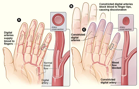
Cold exposure normally causes mild digital vasoconstriction to conserve core heat. In Raynaud phenomenon, this response is exaggerated - digital arteries and arterioles constrict excessively, transiently occluding blood flow to the fingers. As flow returns, characteristic colour changes follow the sequence of ischaemia, deoxygenation and reperfusion.
- White - vasospasm cuts off arterial flow, causing pallor and often numbness
- Blue - stagnant deoxygenated blood in the digits produces cyanosis
- Red - as vasospasm resolves, reactive hyperaemia causes redness, throbbing and pain
Not every patient experiences the full triphasic sequence - white-only or white-then-blue presentations are common, and the classic three-colour description is a helpful pattern to recognise rather than a strict diagnostic requirement.
Clinical features
- Episodic attacks triggered by cold exposure, handling cold objects, or emotional stress, typically lasting minutes to a couple of hours
- Digits most often affected, though the toes, ears, nose and nipples can also be involved
- Numbness or tingling during the white phase, and throbbing pain as blood flow returns in the red phase
- Symptom severity scored by frequency and duration of attacks, and by whether pain or ulceration accompanies them - useful for tracking response to treatment over time
- A careful history of onset age, symmetry, and associated symptoms (skin tightening, joint pain, dry eyes/mouth, dysphagia, breathlessness, photosensitive rash) does most of the diagnostic work at first presentation
Examination
- Inspect the digits between attacks for pitting scars, ulceration, or fixed colour change suggesting chronic ischaemic damage
- Palpate peripheral pulses - normal in Raynaud phenomenon; reduced or absent pulses point toward a large-vessel or embolic cause instead
- Look for skin changes of an underlying connective tissue disease - sclerodactyly, telangiectasia, calcinosis - which may be subtle early on
- Examine the hands for synovitis or joint swelling, and screen more broadly for signs discussed in the individual connective tissue disease articles
Differential diagnosis
- Acrocyanosis - persistent, painless blue discolouration of the hands, without the episodic pattern or pallor phase of Raynaud phenomenon
- Chilblains (perniosis) - localised, itchy, inflamed lesions after cold exposure, rather than transient digital colour change
- Peripheral arterial disease / embolism - a single persistently cold, painful, pulseless digit rather than an episodic bilateral pattern
- Carpal tunnel syndrome - numbness and tingling, but in a median nerve distribution and without colour change
- Erythromelalgia - the near-opposite presentation, with episodic red, hot, burning extremities rather than pallor and cyanosis
Primary versus secondary Raynaud phenomenon
| Feature | Primary (Raynaud disease) | Secondary (Raynaud phenomenon) |
|---|---|---|
| Typical age of onset | Teens to twenties | Over 30 |
| Sex | Strong female predominance | Still commoner in women, less skewed |
| Symmetry | Symmetrical | Often asymmetrical, may affect one hand more |
| Severity | Mild, no tissue damage | Can be severe, with digital ulceration or gangrene |
| Nailfold capillaroscopy | Normal | Abnormal - capillary dilatation and dropout |
| Autoantibodies | Negative | Often positive (ANA, anti-centromere, anti-Scl-70, anti-U1-RNP) |
| Family history | Common | Less relevant - underlying disease drives it |
Causes of secondary Raynaud phenomenon
- Connective tissue disease - systemic sclerosis has by far the strongest association (Raynaud phenomenon occurs in almost all patients, often years before other features), also mixed connective tissue disease, SLE, Sjogren syndrome and dermatomyositis
- Occupational - hand-arm vibration syndrome (vibration white finger) from prolonged use of vibrating tools
- Drugs - beta-blockers, ergotamine, some chemotherapy agents (bleomycin, cisplatin), cocaine and amphetamines
- Haematological - cryoglobulinaemia, polycythaemia, and other hyperviscosity states
- Thoracic outlet syndrome and other causes of vascular compression
- Smoking - a recognised aggravating factor in both primary and secondary disease
Investigations
- Nailfold capillaroscopy - examines the capillaries at the nail bed under magnification; normal in primary disease, showing dilated, distorted or dropped-out capillaries in secondary disease, particularly systemic sclerosis
- ANA and extended autoantibody panel (anti-centromere, anti-Scl-70, anti-U1-RNP, anti-dsDNA) - if secondary disease is suspected clinically
- FBC - polycythaemia or evidence of a haematological cause
- ESR/CRP - raised in an underlying inflammatory connective tissue disease
- Cryoglobulins - if the history suggests a hyperviscosity syndrome
Management
General measures (all patients)
- Keep the whole body, not just the hands, warm - gloves, hand and foot warmers, layered clothing
- Avoid abrupt cold exposure and minimise handling of cold objects (frozen food, cold water)
- Smoking cessation
- Review and, where possible, stop precipitating drugs - particularly beta-blockers
- Avoid excess caffeine, which can worsen vasospasm
Pharmacological treatment
Nifedipine (a dihydropyridine calcium channel blocker) is first-line for patients whose symptoms are not adequately controlled by general measures alone.
- Second-line options - losartan, fluoxetine, or topical glyceryl trinitrate, used where calcium channel blockers are ineffective or not tolerated
- IV prostacyclin analogues (epoprostenol, iloprost) - for severe secondary disease with critical digital ischaemia, given as intermittent infusions
- Endothelin receptor antagonists (bosentan) - reduce the occurrence of new digital ulcers in systemic sclerosis-associated Raynaud phenomenon
- PDE5 inhibitors (sildenafil) - an option for refractory digital ischaemia
- Digital sympathectomy - a surgical option reserved for severe, refractory cases with critical ischaemia threatening tissue loss
Where secondary Raynaud phenomenon is identified, management of the underlying connective tissue disease runs alongside symptomatic treatment of the vasospasm itself.
Complications
Primary Raynaud disease causes discomfort but no lasting tissue damage. Secondary Raynaud phenomenon, particularly in systemic sclerosis, can progress to digital ulceration, chronic pain, and in severe cases critical digital ischaemia and gangrene, occasionally requiring digit amputation. Recurrent digital ulcers are also a source of pain, infection risk and significant impact on hand function and quality of life.
Red flags
Prognosis
Primary Raynaud disease is benign and often improves with simple measures, though it can persist lifelong without causing harm. Secondary Raynaud phenomenon carries the prognosis of its underlying cause - it is a marker of disease rather than a disease in itself, and its severity often, though not always, tracks the severity of the associated connective tissue disease, particularly systemic sclerosis.
References
- NICE Clinical Knowledge Summaries. Raynaud's phenomenon. Available here
- Herrick AL. Raynaud's phenomenon. Journal of Scleroderma and Related Disorders. 2019. Available here
- Belch J, Carlizza A, Carpentier PH et al. ESVM guidelines - the diagnosis and management of Raynaud's phenomenon. VASA. 2017. Available here
- British Society for Rheumatology. Raynaud's phenomenon clinical guidance. Available here
This article is written for revision and education. It is not clinical guidance and must not be used to make decisions about the care of a patient. Always check current NICE guidance and local protocols.