
Typhoid and Enteric Fever
Key points
- Enteric fever: the systemic illness caused by Salmonella enterica serovar Typhi (typhoid) or Paratyphi A, B or C (paratyphoid), spread by the faecal-oral route through contaminated food or water.
- Classic picture: a stepwise, worsening fever over several days, relative bradycardia (Faget sign), headache, abdominal pain and constipation more often than diarrhoea, with faint 'rose spots' on the trunk in a minority.
- A useful clue: relative leukopenia - a normal or low white cell count despite a significant bacterial infection is unusual, and worth noting.
- Complications: intestinal haemorrhage and perforation from necrosis of Peyer's patches, classically in the third week of untreated illness.
- Diagnosis: blood culture is most sensitive early in the illness (bacteraemic phase); stool and urine cultures become more useful later.
- Treatment: ceftriaxone or azithromycin, reflecting widespread fluoroquinolone resistance, particularly in strains from South Asia.
- Chronic carriage: a small proportion become chronic gallbladder carriers, shedding the organism in stool for over a year - the reason for stool clearance testing and work exclusion in food handlers.
Introduction
Enteric fever is a systemic bacterial illness caused by Salmonella enterica serovar Typhi (typhoid fever) or serovars Paratyphi A, B or C (paratyphoid fever), spread by ingesting food or water contaminated with the organism, ultimately traced back to human faecal or urinary shedding. Most UK cases follow travel to South Asia, particularly India, Pakistan and Bangladesh, with further cases linked to parts of Africa and Southeast Asia.
Unlike most Salmonella infections, which cause a self-limiting gastroenteritis confined to the gut (covered in the notes on infective diarrhoea), enteric fever is a systemic, bacteraemic illness - the organism disseminates well beyond the intestine, and diarrhoea is often absent or a late feature rather than the presenting complaint.
Pathophysiology
Ingested organisms survive gastric acid and invade the gut mucosa through M cells overlying the Peyer's patches of the terminal ileum. They are taken up by macrophages, which fail to kill them, and are carried via lymphatics into the bloodstream and on to the reticuloendothelial system - liver, spleen and bone marrow. An initial, often asymptomatic primary bacteraemia is followed, after further multiplication within these organs, by a symptomatic secondary bacteraemia, which is when clinical illness typically begins.
The organism can persist within the gallbladder, particularly in the presence of gallstones, producing chronic carriage with prolonged faecal shedding long after symptoms have resolved - the basis of the historical "Typhoid Mary" cases and the reason public health follow-up continues well beyond clinical recovery.
Clinical features
Untreated, the illness classically evolves over roughly four weeks, though modern presentation is often modified by partial or early treatment.
- Stepwise, progressively rising fever over several days, rather than an abrupt spike
- Relative bradycardia (Faget sign) - the pulse rate is lower than would be expected for the degree of fever, a classically taught but inconsistently present sign
- Headache, malaise and abdominal pain
- Constipation is more typical than diarrhoea, especially early in adults, though diarrhoea can occur and is more common in children and in paratyphoid
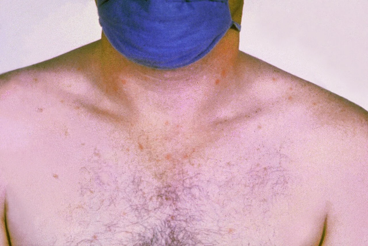
- "Rose spots" - faint, salmon-pink, blanching macules on the trunk, seen in a minority of patients, more visible on lighter skin and easily missed
- Hepatosplenomegaly
- Relative leukopenia - a normal or low white cell count is an unusual finding in significant bacterial infection and a useful supporting clue
Left untreated, illness classically progresses over 4 weeks: a first week of rising fever and bacteraemia; a second week of sustained high fever with rose spots, abdominal tenderness and hepatosplenomegaly; a third week in which the major complications (intestinal haemorrhage, perforation, encephalopathy) appear, reflecting necrosis of the infected Peyer's patches; and, in survivors, a fourth week of gradual convalescence.
Differential diagnosis
- Malaria - the other major cause of fever in a returning traveller, and should always be considered and actively excluded alongside enteric fever rather than instead of it
- Dengue fever - fever, myalgia and thrombocytopenia, with overlapping geographic distribution
- Rickettsial disease - fever with rash, often with an eschar at a tick bite site
- Amoebic liver abscess - fever with right upper quadrant pain
- Acute HIV seroconversion - fever, malaise and rash in a returning traveller with a relevant exposure history
- Brucellosis - undulant fever with an animal or unpasteurised dairy exposure
Investigations
- Blood cultures - the mainstay of diagnosis, most sensitive during the first week (the bacteraemic phase); sensitivity falls as the illness progresses and if antibiotics have already been started
- Bone marrow culture - the single most sensitive test overall, remaining positive even after antibiotics have been started, but invasive and reserved for diagnostically difficult cases
- Stool and urine culture - lower yield early on, but become more useful as the organism is shed later in the illness
- Widal test (agglutination serology) - historically used, but poor sensitivity and specificity limit its value; largely superseded by culture where available, though it may still be used in resource-limited settings
- FBC - relative leukopenia, as above; anaemia and thrombocytopenia can occur
- LFTs - often mildly deranged
Management
Antibiotic choice has changed significantly with the emergence of resistance, and reflects an important, actively examined update to older teaching.
- IV ceftriaxone - first-line empirical treatment, particularly for more unwell patients or where resistance data are unavailable
- Oral azithromycin - an effective alternative for uncomplicated disease
- Treatment duration is typically 7-14 days, guided by clinical response and severity
- Surgical management is required for intestinal perforation, alongside broad-spectrum antibiotics and resuscitation
- Supportive care - adequate fluids and antipyretics; antimotility agents are avoided given the risk of masking or worsening evolving intestinal complications
Public health follow-up
Enteric fever is notifiable and should be reported to UKHSA on clinical suspicion. Because a proportion of patients become chronic carriers, shedding the organism in stool for over a year, stool clearance testing is arranged after treatment, and food handlers and healthcare workers are excluded from work until clearance is confirmed, in line with standard public health protocols for this and other notifiable gastrointestinal infections.
Complications
- Intestinal haemorrhage and perforation - from necrosis of the infected Peyer's patches, typically in the terminal ileum, classically in the second to third week of untreated illness; perforation is a surgical emergency
- Encephalopathy ("typhoid state") - confusion progressing to coma in severe disease
- Myocarditis
- Cholecystitis
- Osteomyelitis - Salmonella is a recognised cause, with a particular association in patients with sickle cell disease
- Chronic carrier state - persistent gallbladder colonisation and faecal shedding, occurring in a small proportion of patients (historically quoted around 1-5%), with important public health implications
Red flags
Prevention
Typhoid vaccination - either an injectable Vi polysaccharide vaccine or an oral live attenuated (Ty21a) vaccine - is recommended for travellers to endemic areas and offers moderate protection (roughly 50-80%, depending on vaccine and setting). Neither vaccine protects against paratyphoid, and vaccination does not replace standard food and water hygiene precautions ("boil it, cook it, peel it, or forget it"), which remain essential regardless of vaccination status.
Prognosis
With prompt, appropriate antibiotic treatment, enteric fever has a good prognosis and most patients recover fully within a few weeks. Delayed diagnosis or treatment - particularly where resistant organisms are treated empirically with an ineffective agent - increases the risk of the third-week complications described above, which is why up-to-date knowledge of local resistance patterns matters as much as recognising the illness itself.
References
- NICE Clinical Knowledge Summaries. Enteric fever (typhoid and paratyphoid). Available here
- UK Health Security Agency. Typhoid and paratyphoid: guidance, data and analysis. Available here
- Parry CM, Hien TT, Dougan G et al. Typhoid fever. New England Journal of Medicine. 2002. Available here
- World Health Organization. Typhoid fact sheet. Available here
This article is written for revision and education. It is not clinical guidance and must not be used to make decisions about the care of a patient. Always check current NICE guidance and local protocols.