
Interpreting the Full Blood Count and Blood Film
Key points
- Work through it systematically: assess each cell line in turn - red cells, white cells, platelets - then ask whether an abnormality is isolated (usually a specific problem) or affects all three (think marrow, sequestration or consumption).
- Classify anaemia by MCV first: microcytic (<80 fL), normocytic (80-100 fL) or macrocytic (>100 fL) - this single number narrows the differential more efficiently than any other.
- Then use the reticulocyte count: in a normocytic anaemia it separates failure of production (low reticulocytes - marrow problem) from loss or destruction (high reticulocytes - bleeding or haemolysis).
- Interpret the white cell differential, not just the total: neutrophilia suggests bacterial infection, steroids or inflammation; lymphocytosis viral infection or CLL; eosinophilia allergy, parasites or drug reaction.
- Always check for artefact: EDTA-induced platelet clumping causes pseudothrombocytopenia, and a delayed or cold sample can distort results. Confirm a surprising result with a blood film before acting on it.
- The film answers questions the analyser cannot: an automated counter reports numbers; the film shows morphology - blasts, schistocytes, sickle cells, hypersegmented neutrophils - which frequently makes the diagnosis outright.
- Urgent film findings: blasts (acute leukaemia), schistocytes with thrombocytopenia (TTP/HUS/DIC), and a leukoerythroblastic film (marrow infiltration) all require same-day discussion with haematology.
- Interpret in clinical context: reference ranges vary with age, sex, ethnicity, altitude and pregnancy, and a result inside the range can still be abnormal for that patient - trends matter more than single values.
Introduction
The full blood count is the most frequently requested test in medicine, and the ability to interpret it methodically - rather than reacting to whichever value the laboratory has flagged - is a core clinical skill. The blood film complements it: where the analyser counts cells, the film looks at them, and morphology often provides the diagnosis directly.1
This article sets out a systematic sequence for reading an FBC, the logic for classifying anaemia, and the blood film findings worth recognising on sight.
A systematic approach
- Check the sample is valid - is it recent, correctly labelled, and consistent with the patient's clinical state? Consider artefact before disease
- Look at the red cells - haemoglobin, then MCV to classify any anaemia, then reticulocytes if normocytic
- Look at the white cells - the total count first, then the differential, which carries most of the information
- Look at the platelets - and if low, exclude clumping on the film before anything else
- Ask: is this one cell line or all three? Isolated abnormalities suggest a specific cause; pancytopenia points to marrow failure, marrow infiltration, sequestration, consumption or severe B12/folate deficiency
- Compare with previous results - a haemoglobin of 105 g/L is reassuring if stable for years and alarming if it was 140 g/L last month
- Interpret in clinical context and request a blood film whenever the picture is unexplained, unexpected or severe
The red cells and classifying anaemia
Anaemia is a reduced haemoglobin concentration for age and sex. The MCV classifies it, and this classification drives the entire subsequent work-up.
| Type | MCV | Common causes |
|---|---|---|
| Microcytic | <80 fL | Iron deficiency (commonest), thalassaemia, anaemia of chronic disease (can be normocytic or microcytic), sideroblastic anaemia, lead poisoning |
| Normocytic | 80-100 fL | Acute blood loss, anaemia of chronic disease, haemolysis, chronic kidney disease (reduced EPO), marrow failure or infiltration, hypothyroidism, pregnancy (dilutional) |
| Macrocytic | >100 fL | B12 or folate deficiency (megaloblastic), alcohol excess, liver disease, hypothyroidism, myelodysplastic syndrome, drugs (methotrexate, hydroxycarbamide, azathioprine), reticulocytosis (reticulocytes are large) |
Using the reticulocyte count
Reticulocytes are immature red cells, and the count reflects how hard the marrow is working. It is most useful in a normocytic anaemia, where it divides the differential cleanly in two:
- High reticulocytes - the marrow is responding appropriately, so red cells are being lost or destroyed: acute bleeding or haemolysis. Look for a raised LDH and unconjugated bilirubin with a low haptoglobin to confirm haemolysis
- Low or inappropriately normal reticulocytes - the marrow is not responding: marrow failure or infiltration, chronic kidney disease, anaemia of chronic disease, or a deficiency state (iron, B12, folate)
Other red cell indices
- MCH (mean corpuscular haemoglobin) - falls alongside MCV in iron deficiency and thalassaemia (hypochromia)
- RDW (red cell distribution width) - a measure of variation in size (anisocytosis). Characteristically raised in iron deficiency but normal in thalassaemia trait, where all the cells are uniformly small - another useful discriminator
- Raised haemoglobin/haematocrit - consider polycythaemia, remembering to distinguish relative (dehydration) from true polycythaemia
The white cells
The differential carries far more information than the total white cell count, which can be normal even when individual lineages are markedly abnormal.
| Abnormality | Common causes |
|---|---|
| Neutrophilia | Bacterial infection, tissue inflammation/necrosis, trauma and surgery, corticosteroids (demargination), myeloproliferative disease, smoking, pregnancy |
| Neutropenia | Chemotherapy (commonest), viral infection, drugs (clozapine, carbimazole, sulfasalazine), marrow failure or infiltration, B12/folate deficiency, autoimmune disease, hypersplenism, ethnic benign neutropenia |
| Lymphocytosis | Viral infection (especially EBV), chronic lymphocytic leukaemia, pertussis, toxoplasmosis, tuberculosis |
| Lymphopenia | Corticosteroids, HIV, severe illness and sepsis, chemotherapy, SLE, lymphoma |
| Eosinophilia | Allergy and atopy (asthma, eczema), parasitic infection, drug reactions, eosinophilic granulomatosis with polyangiitis, adrenal insufficiency, lymphoma, hypereosinophilic syndrome |
| Monocytosis | Chronic infection (tuberculosis), chronic inflammatory disease, chronic myelomonocytic leukaemia, marrow recovery |
| Basophilia | Uncommon and notable - think myeloproliferative neoplasms, particularly CML |
The platelets
- Thrombocytopenia (<150 x10^9/L) - first exclude EDTA-induced platelet clumping on the film (pseudothrombocytopenia), then consider reduced production, increased destruction (ITP, DIC, TTP/HUS, drugs, HIT) or sequestration in hypersplenism
- Thrombocytosis (>450 x10^9/L) - far more often reactive (infection, inflammation, iron deficiency, malignancy, post-splenectomy, bleeding) than clonal (essential thrombocythaemia and other myeloproliferative neoplasms)
- Mean platelet volume (MPV) - large platelets suggest active production (as in ITP) or an inherited platelet disorder such as Bernard-Soulier syndrome
The blood film
The film is examined when the FBC is unexplained, when a specific diagnosis is suspected, or when the analyser flags abnormal cells. Several findings are effectively diagnostic on sight.
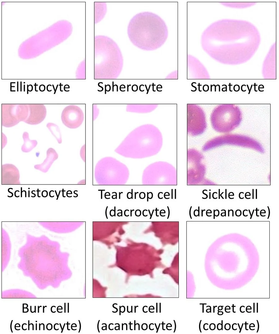
Red cell morphology
| Finding | Description | Associations |
|---|---|---|
| Target cells (codocytes) | Central density surrounded by a pale ring - a "bull's eye" | Thalassaemia, iron deficiency, liver disease, hyposplenism, haemoglobin C disease |
| Spherocytes | Small, dense, round cells lacking central pallor | Hereditary spherocytosis, warm autoimmune haemolytic anaemia |
| Schistocytes | Irregular red cell fragments | Microangiopathic haemolysis - DIC, TTP, HUS, mechanical heart valve, severe pre-eclampsia/HELLP |
| Sickle cells | Elongated, crescent-shaped cells | Sickle cell disease |
| Teardrop cells (dacrocytes) | Pear/teardrop-shaped cells | Myelofibrosis, marrow infiltration, myelodysplasia |
| Pencil cells | Elongated, cigar-shaped hypochromic cells | Iron deficiency anaemia |
| Howell-Jolly bodies | Small round nuclear remnants within red cells | Hyposplenism/asplenia, including functional (sickle cell disease, coeliac disease) |
| Basophilic stippling | Fine blue granules throughout the cytoplasm | Lead poisoning, thalassaemia, sideroblastic anaemia, megaloblastic anaemia |
| Rouleaux | Red cells stacked like a pile of coins | Multiple myeloma, chronic inflammation, very high ESR |
| Heinz bodies and bite cells | Denatured haemoglobin inclusions; cells with a "bite" removed | G6PD deficiency and other oxidative haemolysis |
| Nucleated red blood cells | Immature red cells with retained nuclei | Severe marrow stress, haemolysis, marrow infiltration; normal in neonates |
White cell and other findings
- Blast cells - immature precursors that should not be in the peripheral blood; indicate acute leukaemia and require same-day haematology discussion
- Auer rods - needle-shaped cytoplasmic inclusions confirming myeloid lineage, characteristic of AML
- Hypersegmented neutrophils (6 or more lobes) - B12 or folate deficiency
- Smudge (smear) cells - fragile lymphocytes disrupted during film preparation, classic in CLL
- Atypical/reactive lymphocytes - infectious mononucleosis (EBV) and other viral infections
- Toxic granulation and Dohle bodies - severe bacterial infection and sepsis
- Leukoerythroblastic film - immature white and red cell precursors together in the peripheral blood, indicating marrow infiltration (malignancy, myelofibrosis) or severe marrow stress; always warrants urgent investigation
- Parasites - malaria trophozoites within red cells; the film remains the diagnostic standard
Common artefacts and pitfalls
- EDTA-induced platelet clumping - the analyser reads clumps as single platelets, producing pseudothrombocytopenia in a well patient. Confirmed by clumps on the film and resolved by repeating in a citrate tube
- Delayed or poorly stored samples - cause spuriously raised MCV as cells swell, and haemolysis in transit can affect several parameters
- Very high white cell counts or lipaemia can falsely elevate the measured haemoglobin
- Cold agglutinins cause red cell clumping, giving a spuriously low red cell count and high MCV; warming the sample corrects it
- Dilution from a drip arm - taking the sample from a limb with running IV fluid gives a falsely low haemoglobin; always sample from the opposite arm
- Reference range variation - values differ with age, sex, ethnicity (e.g. benign ethnic neutropenia, in which a lower baseline neutrophil count is normal and does not indicate pathology), altitude and pregnancy (physiological dilutional anaemia and a mild neutrophilia)
Red flags
Putting it together
Interpreting an FBC well is less about memorising reference ranges than about applying a consistent sequence: check the sample, take each cell line in turn, classify any anaemia by MCV and then by reticulocyte count, read the differential rather than the total, compare with previous results, and request a film whenever the picture does not fit.1
Two habits prevent most errors. The first is always considering artefact before disease - clumped platelets, a drip-arm sample or a delayed specimen explain a surprising number of alarming results. The second is treating the patient rather than the number: a result must be interpreted against the clinical presentation, the trend over time, and the patient's own baseline, since a value inside the reference range can still represent significant pathology for that individual.
References
- Bain BJ. Diagnosis from the Blood Smear. N Engl J Med. 2005. Available here
- Bain BJ. Blood Cells: A Practical Guide. Wiley-Blackwell. Available here
- Ed Uthman, S Bhimji and Mikael Häggström, CC BY 4.0, via Wikimedia Commons. Available here
- NICE Clinical Knowledge Summaries (CKS). Anaemia - iron deficiency. 2023. Available here
- British Society for Haematology. Guidelines for the laboratory diagnosis and investigation of blood disorders. Available here
This article is written for revision and education. It is not clinical guidance and must not be used to make decisions about the care of a patient. Always check current NICE guidance and local protocols.