
Antenatal Genetic Screening
Key points
- Screening is not diagnosis: screening gives a chance, not an answer; every high-chance result needs a diagnostic test before any irreversible decision.
- Higher-chance threshold: a chance of 1 in 150 or greater is reported as higher-chance and prompts an offer of further testing.
- Combined test: offered 11+2 to 14+1 weeks - nuchal translucency, PAPP-A and free beta-hCG; around 85% detection for trisomy 21.
- Quadruple test: second-line, 14+2 to 20+0 weeks, screens for trisomy 21 only, with a lower detection rate of around 80%.
- NIPT: cell-free fetal DNA from placental trophoblast; over 99% detection for trisomy 21, but still a screening test offered contingently in the NHS.
- Diagnostic tests: chorionic villus sampling from 11 weeks, amniocentesis from 15 weeks, each with a procedure-related miscarriage risk of around 0.5% or less.
- Haemoglobinopathy screening: offered to all women by 10 weeks for sickle cell and thalassaemia, with partner testing if the woman is a carrier.
- Counselling: must be non-directive and support informed choice, including the choice to decline screening altogether.
Introduction
Antenatal genetic screening identifies pregnancies at increased chance of a genetic or chromosomal condition, so that parents can make informed decisions. In England it is delivered through the NHS Fetal Anomaly Screening Programme (FASP) alongside the sickle cell and thalassaemia screening programme, and every element of it is offered, not imposed - declining is a legitimate and respected choice.1
The single concept underpinning everything here is the distinction between screening and diagnosis. A screening test stratifies a population into higher- and lower-chance groups; it cannot tell an individual woman whether her fetus is affected. A diagnostic test examines fetal genetic material directly and gives a definitive answer, at the cost of a small procedure-related risk of miscarriage. Confusing the two is the commonest error in both exams and clinical practice, and it is the error most likely to cause real harm.
This article covers the screening pathway, the diagnostic tests that follow it, the reproductive options available where a familial condition is already known, and the counselling and ethical framework that surrounds all of it. The conditions themselves are covered in their own articles - Down Syndrome in particular.
What is screened for
- Trisomy 21 (Down syndrome), trisomy 18 (Edwards syndrome) and trisomy 13 (Patau syndrome) - by the combined or quadruple test, and by NIPT where offered
- Sickle cell disease and thalassaemia - offered to all pregnant women in England, ideally by 10 weeks
- Structural fetal anomalies - by the fetal anomaly scan at 18+0 to 20+6 weeks, which screens for eleven specified conditions including anencephaly, open spina bifida, serious cardiac anomalies, bilateral renal agenesis and lethal skeletal dysplasia
- Infectious diseases (HIV, hepatitis B, syphilis) and red cell antibodies - not genetic screening, but part of the same booking bloods and often confused with it
- Specific familial conditions - where a known genetic condition runs in the family, targeted testing is arranged separately through clinical genetics rather than through the population programme
Screening for the trisomies
Screening combines maternal age with biochemical and ultrasound markers to generate a chance - expressed as, for example, 1 in 400. A chance of 1 in 150 or greater is reported as a higher-chance result and prompts the offer of further testing.2
The combined test
Offered between 11+2 and 14+1 weeks, the combined test uses three variables alongside maternal age: nuchal translucency measured on ultrasound, serum pregnancy-associated plasma protein A (PAPP-A), and free beta-hCG. It detects approximately 85% of affected pregnancies for a false positive rate of around 3%.
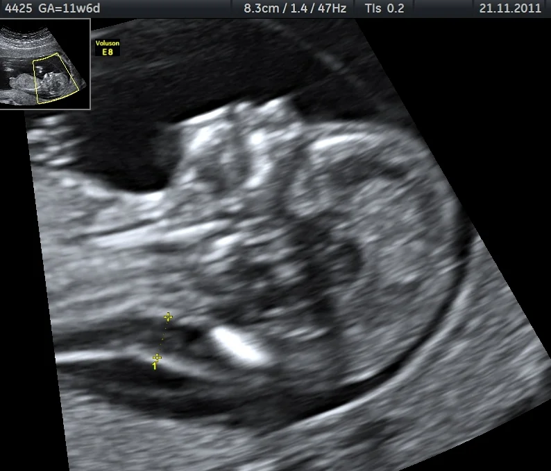
| Condition | Nuchal translucency | PAPP-A | Free beta-hCG |
|---|---|---|---|
| Trisomy 21 (Down) | Increased | Low | High |
| Trisomy 18 (Edwards) | Increased | Low | Low |
| Trisomy 13 (Patau) | Increased | Low | Low |
The quadruple test
If a woman books too late for the combined test, or the nuchal translucency cannot be measured because of fetal position or maternal habitus, the quadruple test is offered between 14+2 and 20+0 weeks. It measures alpha-fetoprotein, unconjugated oestriol, total hCG and inhibin A. In trisomy 21, AFP and oestriol are low while hCG and inhibin A are high. It is a second-line test only, screens for trisomy 21 alone, and has a lower detection rate of around 80%.
Non-invasive prenatal testing (NIPT)
NIPT analyses cell-free fetal DNA in maternal plasma. This DNA derives from placental trophoblast, not the fetus directly, and constitutes roughly 10% of circulating cell-free DNA from around 10 weeks. Its detection rate for trisomy 21 exceeds 99% with a false positive rate below 0.1%, which is why it substantially reduces the number of women proceeding to invasive testing.3
In the NHS it is offered as a contingent second-line test to women with a higher-chance combined or quadruple result, rather than as a universal first-line test - a resource decision, not a reflection of its accuracy. It is also available privately as a first-line test, which means women may present having already had it done.
Diagnostic testing
Definitive diagnosis requires fetal genetic material, obtained by one of two invasive procedures. Both are performed under continuous ultrasound guidance, and both carry a procedure-related miscarriage risk of approximately 0.5% or less in experienced hands - a figure that has fallen considerably from older quoted rates of 1%, and one worth quoting accurately, since it materially affects a woman's decision.
| Chorionic villus sampling | Amniocentesis | |
|---|---|---|
| Timing | From 11 weeks | From 15 weeks |
| Sample | Placental (trophoblast) tissue, transabdominal or transcervical | Amniotic fluid containing fetal cells, transabdominal |
| Advantage | Earlier result, allowing earlier decisions and earlier, safer termination if chosen | Directly fetal in origin, so not subject to confined placental mosaicism |
| Limitation | Confined placental mosaicism can give a discrepant result requiring follow-up amniocentesis | Later in pregnancy; performed before 15 weeks it carries an unacceptable rate of talipes and respiratory morbidity |
| Anti-D | Required if the woman is RhD negative | Required if the woman is RhD negative |
What the laboratory does with the sample
- QF-PCR - rapid aneuploidy testing for chromosomes 13, 18, 21, X and Y, with results within about 3 working days; this is what answers the urgent question
- Full karyotype or chromosomal microarray - takes 2-3 weeks, and detects structural rearrangements and copy number changes beyond the common trisomies
- Targeted single-gene testing - where a specific familial variant is known, arranged in advance through clinical genetics
- Exome sequencing - increasingly used where a structural anomaly is found on scan but karyotype and microarray are normal, through specific NHS pathways
Haemoglobinopathy and carrier screening
Sickle cell disease and thalassaemia are the autosomal recessive conditions screened for universally in pregnancy in England, ideally by 10 weeks, using a combination of the woman's family origin questionnaire and laboratory testing of the booking sample.5
- Screen the mother - full blood count and haemoglobin electrophoresis or HPLC identify carrier states
- If the mother is a carrier, offer testing to the biological father, because the fetus is only at risk if both parents carry a variant
- If both parents are carriers, the fetus has a 1 in 4 chance of being affected, and prenatal diagnosis by CVS or amniocentesis is offered
- Counsel on the options - continuing the pregnancy with the diagnosis known and neonatal care planned, or termination, or declining testing
- Refer to a specialist counsellor in the sickle cell and thalassaemia service, who has the expertise to discuss the specific condition and its prognosis
Reproductive options where a familial condition is known
Where a couple already know they are at risk - because of a previous affected child, a family history, or carrier testing - the decision-making moves earlier, and more options open up. These are discussed through clinical genetics, ideally before conception rather than during an established pregnancy.
- Preimplantation genetic testing (PGT) with IVF - embryos are tested for the specific familial variant and only unaffected embryos transferred, avoiding a decision about an established pregnancy; regulated by the HFEA, which maintains a list of approved conditions
- Prenatal diagnosis by CVS or amniocentesis in a natural conception, with the option of continuing or ending the pregnancy
- Using donor gametes - egg or sperm donation avoids transmitting a variant carried by one partner
- Adoption
- Proceeding without testing - a legitimate choice, sometimes made because the couple would not act on the result, and one that should be supported rather than questioned
- Deciding not to have children - also a legitimate outcome of counselling
For conditions where the familial variant has been identified, the practical prerequisite for almost all of these is that the variant must be known before the pregnancy begins, which is a strong argument for referring couples with a relevant family history pre-conceptually rather than waiting until they present pregnant.
Counselling, consent and ethics
Antenatal screening is ethically distinctive because, unlike most screening, a positive result frequently leads not to treatment but to a decision about whether to continue the pregnancy. That places unusual weight on the quality of consent and the neutrality of the counselling.
Informed choice
- Discussion should happen before the test, early enough to be unhurried, and should cover what is screened for, what a result does and does not mean, and what would follow a higher-chance result
- Declining screening is a valid choice and must be presented as such, not as a failure to engage with care
- Counselling must be non-directive - the clinician's role is to inform, not to steer, and this is particularly important given the profound and personal nature of the decisions involved
- Women should understand that they can accept screening but decline diagnostic testing, or accept diagnostic testing while having already decided to continue the pregnancy regardless - information has value beyond deciding about termination, including preparation and planning delivery in an appropriate unit
- Written information should supplement, not replace, the conversation
Termination for fetal abnormality
Under the Abortion Act 1967, most terminations are performed under ground C before 24 weeks. Ground E permits termination at any gestation, with no upper limit, where there is a substantial risk that the child would be seriously handicapped - the statutory wording, which is now widely regarded as outdated. Two registered medical practitioners must agree. This provision is legally and ethically contested, and disability rights advocates argue that it embodies a discriminatory judgement about the value of disabled lives; students should be able to state the legal position accurately while recognising that it is genuinely disputed rather than settled.4
Delivering a higher-chance or abnormal result
- Deliver in person, in private, promptly, and by someone able to answer the questions that follow
- Use the language of chance rather than 'risk' or 'positive', which carry unhelpful connotations - '1 in 40' is more useful and less alarming than 'high risk'
- Be explicit that a screening result is not a diagnosis, and explain what the confirmatory options are
- Allow time - there is almost never a need to decide about invasive testing the same day
- Offer referral to fetal medicine, clinical genetics and, where relevant, condition-specific support organisations
- Both parents should be present where the woman wishes it, and interpreters used where needed rather than relying on family members
After the pregnancy
Screening does not stop at delivery. The newborn blood spot (heel-prick) test at around day 5 screens for nine conditions including phenylketonuria, congenital hypothyroidism, sickle cell disease, cystic fibrosis and MCADD, and the newborn hearing screen and the newborn and infant physical examination follow shortly after. A normal antenatal screening pathway does not remove the need for any of these, and parents should understand that the two programmes look for different things.
Where a genetic condition is confirmed, whether antenatally or postnatally, the family should be referred to clinical genetics for full counselling, accurate recurrence risk for future pregnancies, and consideration of cascade testing for relatives - the point at which one pregnancy's screening result becomes relevant to a whole family.
References
- NHS England. Fetal anomaly screening programme (FASP) handbook. Available here
- NICE NG201. Antenatal care. 2021. Available here
- Royal College of Obstetricians and Gynaecologists. Non-invasive prenatal testing for chromosomal abnormality using maternal plasma DNA. Scientific Impact Paper No. 15. Available here
- Royal College of Obstetricians and Gynaecologists. Termination of pregnancy for fetal abnormality in England, Scotland and Wales. Available here
- NHS England. Sickle cell and thalassaemia screening programme handbook. Available here
This article is written for revision and education. It is not clinical guidance and must not be used to make decisions about the care of a patient. Always check current NICE guidance and local protocols.