
Malignant Melanoma
Key points
- Malignant melanoma: a malignant tumour of melanocytes, and the most dangerous form of skin cancer because of its propensity to metastasise early, even from a primary lesion that is still small.
- Main risk factor: intermittent, intense UV exposure and episodes of sunburn - particularly in childhood - correlate more strongly with melanoma risk than cumulative lifetime sun exposure, unlike basal cell and squamous cell carcinoma.
- Recognition: the ABCDE criteria (Asymmetry, Border irregularity, Colour variation, Diameter over 6mm, Evolution) and the weighted Glasgow 7-point checklist both help identify a suspicious pigmented lesion needing urgent referral.
- Subtypes: superficial spreading (commonest in the UK), nodular (most aggressive, vertical growth from the outset), lentigo maligna melanoma (elderly, chronically sun-damaged skin), and acral lentiginous (palms, soles, nail bed - not UV-related).
- Breslow thickness: the single most important prognostic factor - the depth of invasion in millimetres from the granular layer to the deepest tumour cell - and the main determinant of excision margin and further staging.
- Diagnosis: complete excision biopsy with a narrow (2mm) margin, never an incisional, shave or punch biopsy, since accurate Breslow thickness measurement on the whole lesion is essential.
- Management: wide local re-excision with a margin determined by Breslow thickness, sentinel lymph node biopsy for intermediate/high-risk disease, and adjuvant or systemic immunotherapy or targeted therapy for high-risk or metastatic disease.
- Red flag: any new or changing pigmented lesion meeting ABCDE or 7-point checklist criteria needs urgent (2-week-wait) referral rather than reassurance or watchful waiting in primary care.
Introduction
Malignant melanoma is a cancer arising from melanocytes, the pigment-producing cells of the epidermis. It accounts for a small minority of skin cancers overall but causes the large majority of skin cancer deaths, because of its distinctive tendency to metastasise via both the lymphatic and haematogenous routes at a relatively early stage, sometimes from a primary lesion only a few millimetres thick.3
Incidence has risen steadily in the UK over recent decades, largely attributed to changing patterns of sun exposure and holiday travel, though prognosis for early-detected disease is excellent. The entire clinical emphasis in this topic is therefore on early recognition - correctly identifying a suspicious pigmented lesion before it has invaded deeply - since this single step does more for outcome than any treatment given after the fact.
Risk factors
- Ultraviolet exposure - particularly intermittent, intense exposure and episodes of sunburn, especially in childhood; this pattern correlates more strongly with melanoma than the cumulative, occupational sun exposure more typical of basal cell and squamous cell carcinoma
- Fitzpatrick skin type I-II - fair skin, red or blond hair, light eyes, and a tendency to burn rather than tan
- High naevus count - more than 100 common naevi, or the presence of atypical (dysplastic) naevi
- Personal or family history of melanoma - a first-degree relative with melanoma roughly doubles risk; germline CDKN2A mutations account for a proportion of familial melanoma
- Immunosuppression - organ transplant recipients and other immunosuppressed patients have an increased incidence and often a more aggressive course
- Sunbed use, particularly starting before age 35
- Previous melanoma - a strong risk factor for a second, separate primary melanoma
Pathophysiology and classification
Melanoma arises from malignant transformation of melanocytes, usually driven by cumulative UV-induced DNA damage in a genetically susceptible individual. Early growth is typically confined to the epidermis and superficial dermis, spreading outwards (radial growth phase), before a subset of cells acquire the capacity to invade deeper into the dermis (vertical growth phase), which is the point at which metastatic potential increases sharply - the rationale behind Breslow thickness as the key prognostic measurement.
| Subtype | Features |
|---|---|
| Superficial spreading | The commonest subtype in fair-skinned populations (around 70% of UK melanomas); a flat or slightly raised, irregular, variably pigmented lesion with a prolonged radial growth phase before vertical invasion |
| Nodular | The second commonest and most aggressive subtype; a rapidly growing, often uniformly dark (or amelanotic) nodule that is vertically invasive from the outset, without a preceding radial growth phase, meaning ABCDE criteria are less reliable and a nodule that simply grows and bleeds should still raise concern |
| Lentigo maligna melanoma | Arises within a pre-existing lentigo maligna (melanoma in situ) on chronically sun-damaged skin, typically the face, in older patients; grows slowly over years before invasion |
| Acral lentiginous | Occurs on the palms, soles, and beneath the nail (subungual); not associated with UV exposure, and is the relatively commonest subtype in patients with darker skin, where overall melanoma incidence is much lower |
Clinical features
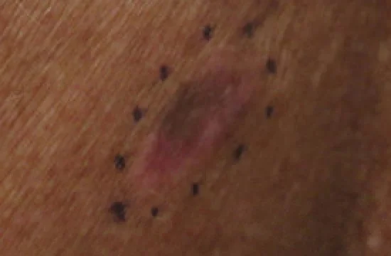
The ABCDE criteria
| Feature | What to look for |
|---|---|
| Asymmetry | One half of the lesion does not match the other |
| Border irregularity | Edges are ragged, notched or blurred rather than smooth |
| Colour variation | More than one shade - tan, brown, black, and sometimes red, white or blue - within a single lesion |
| Diameter | Greater than 6mm, though melanoma can be diagnosed at a smaller size |
| Evolution | Any change in size, shape, colour, or new symptoms (itch, bleeding) over weeks to months |
The Glasgow 7-point checklist
Used in the UK alongside or instead of ABCDE, this weighted checklist separates major features (each scoring 2 points) from minor features (each scoring 1 point); a score of 3 or more prompts referral.6
- Major features (2 points each) - change in size, change in shape, change in colour
- Minor features (1 point each) - diameter 7mm or more, inflammation, oozing or bleeding, altered sensation (itch)
Amelanotic melanoma lacks the pigmentation of typical melanoma and presents instead as a pink, red or skin-coloured nodule or patch, which can easily be mistaken for a pyogenic granuloma, basal cell carcinoma or simple inflammatory lesion - a genuine diagnostic trap that accounts for some of the delayed diagnoses seen in practice.
Clinical examination
- Full skin examination, since patients (and clinicians) often focus on the lesion of concern and miss a second, separate primary elsewhere
- Dermoscopy - increasingly used in primary and secondary care to improve diagnostic accuracy beyond the naked eye, looking for an atypical pigment network, irregular streaks, blue-white structures and atypical vascular patterns
- Regional lymph node examination - the nodal basin draining the site of the lesion should always be examined for palpable lymphadenopathy
- Photography and measurement - documented size and appearance at baseline assists in judging genuine evolution at subsequent review
- Examination of the nails, palms and soles - easily overlooked sites for acral lentiginous melanoma
Differential diagnosis
- Benign melanocytic naevus - stable over years, symmetrical, evenly pigmented; see Benign Skin Lesions for the normal life cycle of a naevus
- Seborrhoeic keratosis - a 'stuck-on', warty surface with horn cysts on dermoscopy, rather than a pigment network
- Dermatofibroma - a firm dermal nodule with a positive dimple sign on lateral compression
- Pyogenic granuloma - a rapidly growing, friable, bleeding vascular lesion that can closely mimic amelanotic nodular melanoma and should be excised for histology if there is any doubt
- Subungual haematoma - a pigmented band from trauma that grows out with the nail and does not widen proximally, unlike subungual melanoma
- Basal cell carcinoma (pigmented subtype) - a pearly, rolled edge with telangiectasia rather than the irregular pigment network of melanoma
Investigations
Any lesion suspicious for melanoma should be managed with a clear diagnostic pathway rather than piecemeal sampling.
- Complete excision biopsy with a narrow (around 2mm) margin and a cuff of subcutaneous fat is the correct initial procedure - never an incisional, punch or shave biopsy, since accurate Breslow thickness (and therefore prognosis and further management) depends on measuring the whole lesion
- Histopathology reports Breslow thickness, presence or absence of ulceration, mitotic rate, and margin clearance, all of which feed directly into staging and the decision for re-excision margin
- Sentinel lymph node biopsy - offered to patients with Breslow thickness over 1mm (or over 0.8mm with ulceration), to stage the regional lymph node basin without the morbidity of a full lymph node dissection1
- Staging CT or PET-CT - considered for higher-stage disease (thick primary, positive sentinel node, or clinical suspicion of metastasis) to look for nodal or distant spread
- BRAF mutation testing - performed on advanced or metastatic melanoma tissue to determine eligibility for targeted BRAF/MEK inhibitor therapy
Management
Wide local excision
Once Breslow thickness is known from the initial excision biopsy, a wide local re-excision is performed with a margin determined by thickness, to reduce the risk of local recurrence.8
| Breslow thickness | Excision margin |
|---|---|
| In situ (confined to the epidermis) | 5mm |
| Up to 1mm | 1cm |
| 1.01-2mm | 1-2cm |
| 2.01-4mm | 2-3cm |
| Over 4mm | 2-3cm |
Nodal and systemic management
- Sentinel lymph node biopsy as above, to stage regional nodes for intermediate/high-risk disease; a positive sentinel node informs prognosis and the discussion around adjuvant therapy, though completion lymph node dissection is no longer routinely performed for every positive sentinel node given trial evidence of limited benefit
- Adjuvant systemic therapy - anti-PD-1 immunotherapy (nivolumab, pembrolizumab) or targeted BRAF/MEK inhibitor therapy (for BRAF-mutant disease) is now offered to patients with high-risk resected disease (positive nodes, or thick/ulcerated primaries), reducing recurrence risk
- Metastatic disease - immune checkpoint inhibitors, alone or in combination (nivolumab plus ipilimumab), and targeted BRAF/MEK inhibitor combinations for BRAF-mutant disease, have transformed outcomes in advanced melanoma over the past decade, with durable long-term responses seen in a meaningful proportion of patients who would previously have had a very short life expectancy7
- Radiotherapy - used for symptom palliation in metastatic disease, or as adjuvant treatment to a nodal basin in selected cases
Follow-up
Structured follow-up with clinical skin and nodal examination is offered for a period after diagnosis, with the frequency and duration depending on stage, to detect local recurrence, in-transit metastasis, or a new primary melanoma early, alongside patient education on sun protection and self-examination.
Complications
- Local recurrence at the excision site
- In-transit metastasis - tumour deposits in the skin or subcutaneous tissue between the primary site and the regional lymph node basin
- Regional lymph node metastasis
- Distant metastasis - melanoma can spread almost anywhere, but has a particular tendency for the lungs, liver, brain, bone and, distinctively, the small bowel and other unusual visceral sites
- Psychological impact - considerable anxiety around recurrence risk and the need for ongoing surveillance, particularly in the first few years after diagnosis
Red flags
Prognosis
Prognosis in melanoma is overwhelmingly determined by Breslow thickness and stage at diagnosis rather than subtype, which is why early recognition dominates the clinical approach to this disease.4,5
| Stage | Approximate 5-year survival |
|---|---|
| Stage I (thin, node-negative) | Over 95% |
| Stage II (thicker, node-negative) | Roughly 80-90% |
| Stage III (regional node involvement) | Roughly 40-70%, depending on nodal burden |
| Stage IV (distant metastasis) | Historically under 20%, though modern immunotherapy and targeted therapy have meaningfully improved outcomes for many patients |
The steep fall in survival between early, thin, node-negative disease and later, thicker or node-positive disease is the single most important prognostic fact in this topic, and is the reason melanoma is fundamentally a disease where the greatest gains come from recognising it early rather than from treating it once advanced - though the systemic treatments now available for metastatic disease have genuinely changed what was, within living memory, a near-uniformly fatal diagnosis at that stage.
References
- NICE NG14. Melanoma: assessment and management. 2015. Available here
- NICE NG12. Suspected cancer: recognition and referral. 2015, updated 2023. Available here
- Cancer Research UK. Melanoma skin cancer statistics. Available here
- Gershenwald JE, Scolyer RA, Hess KR et al. Melanoma staging: evidence-based changes in the American Joint Committee on Cancer eighth edition cancer staging manual. CA: A Cancer Journal for Clinicians. 2017. Available here
- Breslow A. Thickness, cross-sectional areas and depth of invasion in the prognosis of cutaneous melanoma. Annals of Surgery. 1970. Available here
- MacKie RM. Clinical recognition of early invasive malignant melanoma. BMJ. 1990. Available here
- Larkin J, Chiarion-Sileni V, Gonzalez R et al. Combined nivolumab and ipilimumab or monotherapy in untreated melanoma. New England Journal of Medicine. 2015. Available here
- Marsden JR, Newton-Bishop JA, Burrows L et al. Revised UK guidelines for the management of cutaneous melanoma 2010. British Journal of Dermatology. 2010. Available here
This article is written for revision and education. It is not clinical guidance and must not be used to make decisions about the care of a patient. Always check current NICE guidance and local protocols.