
Benign Skin Lesions
Key points
- Why this matters: correctly recognising a benign skin lesion clinically avoids unnecessary excision and patient anxiety, while still catching the minority of lesions that genuinely need referral for a tissue diagnosis.
- Seborrhoeic keratosis: the commonest benign epidermal tumour of older adults - a well-demarcated, 'stuck-on', waxy or warty papule or plaque, often pigmented, requiring no treatment unless symptomatic or diagnostically uncertain.
- Melanocytic naevi: benign proliferations of melanocytes; new naevi commonly appear throughout childhood and adolescence and the total number stabilises in early adulthood, after which a new or changing naevi needs assessment.
- Dermatofibroma: a firm dermal nodule, often following minor trauma or an insect bite, with a pathognomonic dimple (Fitzpatrick) sign - central depression on lateral compression.
- Skin tags (acrochordons): soft, pedunculated, flesh-coloured papules in flexures, strongly associated with obesity, insulin resistance and pregnancy.
- Epidermoid and pilar cysts: firm, mobile, subcutaneous nodules; epidermoid cysts typically show a central punctum and favour the face and trunk, while pilar cysts arise on the scalp and usually lack one.
- Vascular lesions: cherry angiomas (Campbell de Morgan spots) are common, harmless, and increase with age; pyogenic granuloma is a rapidly growing, easily bleeding vascular lesion that can mimic amelanotic melanoma.
- When to worry: a changing, growing, bleeding or non-healing lesion - especially a pigmented one - needs assessment against the 2-week-wait skin cancer referral criteria rather than being assumed benign.
Introduction
Benign skin lesions are extremely common, and the great majority of lumps and bumps referred from primary care, or noticed incidentally on examination, fall into a small number of well-defined, easily recognised categories. Confident clinical recognition matters for two reasons: it avoids unnecessary excision, biopsy and patient anxiety over lesions that need no treatment, and it sharpens the ability to spot the minority of lesions that do not fit a benign pattern and genuinely need referral.
This note covers the lesions most often encountered and tested: seborrhoeic keratosis, melanocytic naevi, dermatofibroma, skin tags, epidermoid and pilar cysts, and common benign vascular lesions. Lipomas are covered separately, and malignant skin lesions - melanoma, basal cell carcinoma and squamous cell carcinoma - each have their own dedicated notes; this article focuses on telling benign lesions apart from each other and knowing when a lesion has stepped outside a reassuring pattern.
Seborrhoeic keratosis
Seborrhoeic keratosis is the commonest benign epidermal tumour, arising from proliferation of epidermal keratinocytes. It is extremely common from middle age onwards, increasing in number with age, and has no malignant potential.1
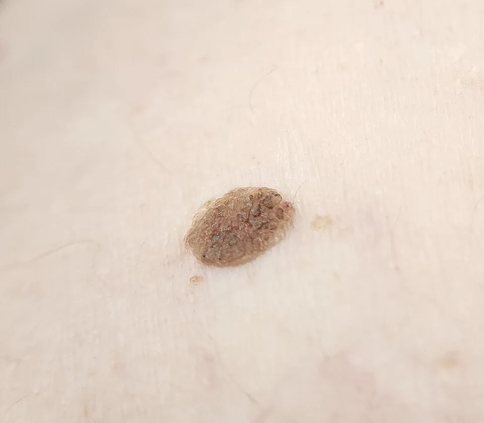
Lesions are well-demarcated, raised, and classically described as looking 'stuck on' to the skin surface, with a warty, greasy or waxy texture and a colour ranging from skin-coloured to light brown to almost black. They vary from a few millimetres to several centimetres, occur anywhere except the palms, soles and mucous membranes, and are often multiple. Dermoscopy characteristically shows horn (milia-like) cysts and comedone-like openings, which help confirm the diagnosis when it is not obvious clinically.
No treatment is needed unless a lesion is symptomatic (catching on clothing, itching) or the patient requests removal for cosmetic reasons, or if there is diagnostic uncertainty. Cryotherapy, curettage or shave excision are effective options where removal is wanted.
Melanocytic naevi
A melanocytic naevus (mole) is a benign proliferation of melanocytes, and most people accumulate a variable number - typically 20-40 - throughout childhood and adolescence, with the total stabilising in early adulthood.2
| Type | Features |
|---|---|
| Junctional | Melanocytes at the dermo-epidermal junction; flat, evenly pigmented macule, often the earliest stage |
| Compound | Melanocytes at the junction and within the dermis; a raised, pigmented papule, often with a smoother surface |
| Intradermal | Melanocytes confined to the dermis; a raised, often paler or skin-coloured papule, typically seen in older adults as a naevus matures and loses surface pigment |
This progression - flat and pigmented in youth, becoming raised and paler with age - is the normal life cycle of an individual naevus and is not itself concerning. What matters clinically is change that is asymmetric, rapid, or occurs in a naevus that had previously been stable for years, particularly in adulthood, since new naevi appearing for the first time after around age 40, or any naevus changing outside this predictable pattern, warrant assessment.
Atypical (dysplastic) naevi are larger, less symmetrical, and more variably pigmented than a typical naevus, and patients with multiple atypical naevi carry a higher lifetime melanoma risk and benefit from surveillance. Recognising a naevus that has developed genuinely melanoma-suspicious features - asymmetry, an irregular border, colour variation, a diameter over 6mm, or evolution - is covered in detail in the article on malignant melanoma; any naevus with these features should follow the same 2-week-wait pathway as a new pigmented lesion.
Dermatofibroma
A dermatofibroma (also called a fibrous histiocytoma) is a firm, benign dermal nodule composed of fibroblasts and histiocytes, thought in many cases to represent an exaggerated reactive response to minor trauma such as an insect bite or a shaving nick.3
It presents as a firm, slow-growing, dome-shaped or slightly depressed papule or nodule, usually a few millimetres to a centimetre across, most often on the lower legs, and typically pigmented tan to brown, though colour is variable. The lesion is fixed to the overlying skin but mobile over deeper structures.
No treatment is required unless the lesion is symptomatic or the diagnosis is uncertain, in which case excision provides both treatment and histological confirmation.
Skin tags (acrochordons)
Skin tags are soft, benign, pedunculated papules of skin-coloured or slightly hyperpigmented tissue, typically a few millimetres long, found in flexural sites - the neck, axillae, groin and eyelids - where skin rubs against skin or clothing.4
They are strongly associated with obesity, insulin resistance and type 2 diabetes, and commonly increase in number during pregnancy, reflecting hormonal and metabolic influences on skin growth. They are entirely benign and asymptomatic other than mechanical irritation from catching on clothing or jewellery, or occasional torsion causing acute painful thrombosis of a tag.
Removal (snip excision, cryotherapy, or cautery) is offered only for symptomatic or cosmetically bothersome lesions and is not medically necessary.
Epidermoid and pilar cysts
Epidermoid cysts (often loosely called sebaceous cysts, though they do not arise from sebaceous glands) are the commonest cutaneous cyst, formed from a proliferation of epidermis within the dermis, often following occlusion of a hair follicle. They present as firm, round, mobile, subcutaneous nodules, most often on the face, neck, and trunk, frequently with a visible central punctum - a small, often blocked, dark or skin-coloured pore at the surface - through which cheesy, keratinaceous material can sometimes be expressed.5
Pilar (trichilemmal) cysts are histologically distinct but clinically similar, arising instead from the hair follicle outer root sheath; they occur almost exclusively on the scalp, are often multiple and familial, and characteristically lack a punctum, which is a useful distinguishing clue.
Both types can become inflamed or secondarily infected, presenting as a tender, erythematous, sometimes discharging swelling - which needs treating as an abscess (incision and drainage, with antibiotics if there is surrounding cellulitis) rather than attempting definitive cyst wall excision in the acute setting. Elective complete excision of the cyst wall, once any inflammation has settled, is the only way to prevent recurrence, since incision and drainage alone leaves the cyst wall in place.
Vascular lesions
Cherry angioma (Campbell de Morgan spots)
Small, bright red or purple, dome-shaped papules composed of dilated capillaries, extremely common from middle age onwards and increasing steadily in number with age. They are entirely benign, asymptomatic, and require no treatment beyond reassurance; removal (if requested for cosmetic reasons) can be achieved with laser or electrocautery.
Pyogenic granuloma
A rapidly growing, friable, bright red or purple vascular nodule that bleeds easily with minor trauma, often appearing over days to a few weeks, classically following minor trauma and with a particular tendency to occur during pregnancy (sometimes called a granuloma gravidarum, often on the gums) or in association with certain medications (retinoids, some antiretrovirals).6
Treatment is with curettage and cautery, or shave excision, both of which also provide a histological specimen; lesions can recur, particularly if the base is not adequately treated.
Milia
Tiny (1-2mm), firm, white or yellow superficial keratin-filled cysts, extremely common on the face, particularly around the eyelids, and in neonates. They are entirely benign, need no treatment, and can be simply incised and expressed if removal is requested.
Clinical examination
- Full history - duration, rate of change, symptoms (itch, bleeding, pain), and any preceding trauma
- Site, size, colour, surface texture and border - a 'stuck-on' warty surface, a dimple sign on compression, and a visible punctum are each highly specific bedside findings for their respective diagnoses
- Mobility - fixed to skin but mobile over deeper tissue (cyst, dermatofibroma) versus fixed to deeper structures
- Dermoscopy - can confirm a seborrhoeic keratosis (horn cysts) or a vascular lesion, and is increasingly used to support (though never replace) clinical judgement in pigmented lesions
- Rate of change since the last review, if the lesion has been seen before, since this is often more informative than a single snapshot assessment
When to refer
Most benign skin lesions need no referral at all. Referral under the 2-week-wait suspected skin cancer pathway is appropriate for any lesion with features suggesting melanoma, basal cell carcinoma or squamous cell carcinoma, detailed in their respective articles, but broadly includes:7
- A new or changing pigmented lesion with asymmetry, border irregularity, colour variation, a diameter over 6mm, or evolution over weeks to months
- A non-healing lesion, or one that bleeds or ulcerates without a clear benign explanation
- A rapidly growing lesion, particularly in an older patient, without a convincing benign cause such as recent trauma
- Any lesion where the patient, or the examining clinician, remains concerned despite an apparently reassuring appearance
Complications
- Secondary infection of an inflamed epidermoid or pilar cyst
- Bleeding from a traumatised dermatofibroma, skin tag or pyogenic granuloma
- Scarring from excision, particularly relevant when counselling patients about purely cosmetic removal of an entirely benign lesion
- Diagnostic delay if an atypical-looking benign lesion is removed without histology and later recurs or is later found to have been misdiagnosed
- Unnecessary anxiety from an incorrectly labelled benign lesion, or conversely false reassurance about a lesion that in fact needed referral
Red flags
Prognosis
All of the lesions covered here are benign and, once correctly identified, carry an excellent prognosis with no treatment needed beyond reassurance, unless removal is wanted for symptomatic or cosmetic reasons. Seborrhoeic keratoses and cherry angiomas simply accumulate gradually with age; dermatofibromas, skin tags, cysts and milia are generally stable once established.
The main risk in this area of practice is not under-treating a benign lesion but misclassifying an early malignant lesion as benign, which is why the emphasis throughout is on recognising the specific, reassuring features of each benign diagnosis - the dimple sign, the visible punctum, the stuck-on surface - rather than simply excluding malignancy by a lesion's absence of obvious alarm features. When a lesion does not cleanly fit one of these recognisable patterns, biopsy or excision for histology, or referral, is the safer course.
References
- DermNet NZ. Seborrhoeic keratosis. Available here
- DermNet NZ. Melanocytic naevus. Available here
- DermNet NZ. Dermatofibroma. Available here
- DermNet NZ. Skin tag. Available here
- DermNet NZ. Epidermoid cyst. Available here
- DermNet NZ. Pyogenic granuloma. Available here
- NICE NG12. Suspected cancer: recognition and referral. 2015, updated 2023. Available here
- Schwartz RA. Sign of Leser-Trelat. Journal of the American Academy of Dermatology. 1996. Available here
This article is written for revision and education. It is not clinical guidance and must not be used to make decisions about the care of a patient. Always check current NICE guidance and local protocols.