
Basal Cell Carcinoma
Key points
- Basal cell carcinoma: the commonest skin cancer and commonest cancer of any kind in the UK, a locally invasive tumour of basal keratinocytes that grows slowly and almost never metastasises.
- Main risk factor: cumulative lifetime UV exposure, in contrast to the intermittent, intense sunburn pattern more strongly linked to melanoma - which is why BCC favours chronically sun-exposed sites, especially the head and neck.
- Nodular BCC: the commonest subtype - a pearly, rolled-edge papule or nodule with visible telangiectasia, which can ulcerate centrally (the classically described 'rodent ulcer').
- Other subtypes: superficial (a scaly, erythematous patch on the trunk with a thin rolled edge) and morphoeic/sclerosing (a scar-like, ill-defined, more aggressive plaque with a higher recurrence rate).
- Diagnosis: clinical, supported by dermoscopy; unlike melanoma, an incisional, punch or shave biopsy is acceptable to confirm subtype before planning definitive treatment.
- Management: surgical excision with a margin matched to risk, Mohs micrographic surgery for high-risk or recurrent lesions and cosmetically sensitive sites, and topical treatment or cryotherapy for low-risk superficial disease.
- Gorlin syndrome: an autosomal dominant condition (PTCH1 mutation) causing multiple BCCs from a young age, odontogenic jaw cysts and skeletal abnormalities.
- Prognosis: excellent, with near-zero mortality, but neglected or high-risk lesions can invade deeply into cartilage, bone or the orbit if left untreated - hence the historical name 'rodent ulcer'.
Introduction
Basal cell carcinoma (BCC) is a locally invasive malignant tumour of basal keratinocytes, and is the commonest cancer of any type diagnosed in the UK, though it is often excluded from standard cancer statistics because it is so rarely fatal. Incidence rises steadily with age and cumulative sun exposure, and it is now also seen with increasing frequency in younger adults.3
The clinical priority with BCC is quite different from melanoma: rather than racing to prevent early metastasis, which is vanishingly rare, the goal is complete local clearance to prevent progressive, disfiguring local tissue destruction - historically severe enough to erode through cartilage and bone, which is where the old term 'rodent ulcer' comes from.2
Risk factors
- Cumulative lifetime UV exposure - the dominant risk factor, correlating with total sun exposure over decades rather than episodes of sunburn, which is why BCC is so strongly associated with chronic occupational or recreational sun exposure on the head, neck and forearms
- Fitzpatrick skin type I-II - fair skin, a tendency to burn rather than tan
- Increasing age
- Immunosuppression - organ transplant recipients have a markedly increased risk, though relatively more so for squamous cell carcinoma
- Previous BCC - a strong predictor of further lesions, often at a different site
- Ionising radiation exposure, including previous radiotherapy
- Arsenic exposure
- Gorlin syndrome (naevoid basal cell carcinoma syndrome) - an autosomal dominant condition causing multiple BCCs from a young age, discussed further below
Pathophysiology
BCC arises from basal keratinocytes of the epidermis and hair follicle, driven in the great majority of cases by aberrant activation of the Hedgehog signalling pathway. Loss-of-function mutations in PTCH1 (which normally inhibits the pathway) or activating mutations in SMO (Smoothened, a downstream signal transducer) remove the normal brake on Hedgehog signalling, driving uncontrolled basal cell proliferation.4
This mechanistic understanding has direct treatment relevance: the Hedgehog pathway inhibitors vismodegib and sonidegib, used in advanced or metastatic disease, work by directly blocking SMO, restoring the normal brake on this signalling pathway.5
Clinical features
BCC occurs predominantly on chronically sun-exposed skin, especially the head and neck (particularly the nose, forehead and periorbital area), though it can occur on the trunk and limbs too.
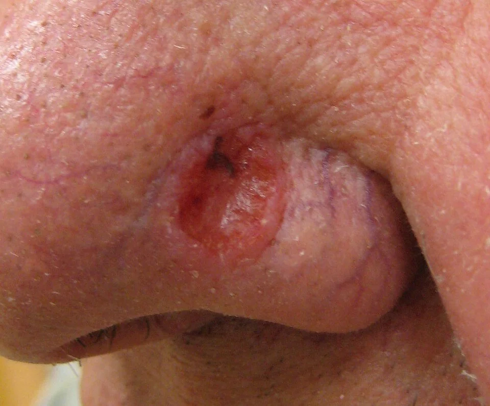
| Subtype | Features |
|---|---|
| Nodular (commonest) | A pearly, translucent papule or nodule with a rolled, well-defined edge and visible surface telangiectasia; can ulcerate centrally, historically called a 'rodent ulcer' |
| Superficial | A well-demarcated, scaly, erythematous patch or thin plaque, often on the trunk, with a fine, thread-like rolled edge best seen on stretching the skin; can be mistaken for eczema or psoriasis |
| Morphoeic (sclerosing) | An ill-defined, waxy, scar-like plaque without a clear rolled edge; more locally aggressive, infiltrative, and has the highest recurrence rate of the common subtypes |
| Pigmented | Any of the above patterns with brown or black pigmentation, which can cause confusion with melanoma |
| Basosquamous | A mixed histological pattern with features of both BCC and squamous cell carcinoma, behaving somewhat more aggressively than a pure BCC |
Growth is characteristically slow, over months to years, and lesions are usually asymptomatic other than occasional bleeding or crusting from minor trauma, which is a common reason patients present ('a spot that won't heal' or 'keeps bleeding when I shave').
Clinical examination
- Site, size and subtype-defining features - rolled edge, telangiectasia, ulceration, or the ill-defined scar-like quality of a morphoeic lesion
- Dermoscopy - characteristically shows arborising (branching) telangiectatic vessels, blue-grey ovoid nests, and an absence of a pigment network, all supporting the diagnosis
- Assessment of high-risk features - size, site (particularly the 'H-zone' of the face - central face, eyelids, eyebrows, periorbital, nose, lips, chin, ear, temple), poorly defined margins, and recurrent disease, all of which influence the choice between standard excision and Mohs micrographic surgery
- Full skin examination - patients with one BCC are at increased risk of further lesions at other sites
Differential diagnosis
- Squamous cell carcinoma - typically more keratotic, crusted or ulcerated, and can arise from a pre-existing actinic keratosis; see Squamous Cell Carcinoma and Bowen Disease
- Actinic keratosis - a rough, scaly, erythematous patch on sun-damaged skin, generally flatter and without a rolled edge
- Seborrhoeic keratosis - a 'stuck-on', warty surface with horn cysts on dermoscopy, lacking telangiectasia or a rolled edge
- Molluscum contagiosum - dome-shaped, umbilicated papules, generally in children or immunosuppressed adults rather than the older, sun-damaged skin typical of BCC
- Pigmented BCC versus melanoma - dermoscopy is particularly useful here, since pigmented BCC lacks a true pigment network and instead shows blue-grey ovoid nests and arborising vessels
- Psoriasis or eczema - for superficial BCC on the trunk, which can be mistaken for an inflammatory patch that simply fails to respond to topical steroids
Investigations
Diagnosis is usually clinical, supported by dermoscopy, with biopsy used to confirm the diagnosis and subtype before planning definitive treatment.
- Biopsy (incisional, punch or shave) - unlike a suspected melanoma, a partial biopsy is acceptable for BCC, since accurate subtyping (rather than a single thickness measurement) is what guides management, and BCC does not carry the same staging implications from incomplete initial sampling
- Dermoscopy - arborising vessels, blue-grey ovoid nests, and absence of a pigment network support the diagnosis and can help distinguish subtype
- Imaging (CT or MRI) - reserved for large, neglected, or deeply invasive lesions where bone, cartilage or orbital invasion is suspected, particularly around the nose, ear or eye
Management
Treatment is chosen according to subtype, site and risk of recurrence, since the great majority of BCCs are curable with the correct initial approach.1
Surgical excision
Standard surgical excision with a 4mm margin clears the great majority of low-risk, well-defined BCCs. Higher-risk lesions (morphoeic subtype, recurrent disease, poorly defined margins, or those in the high-risk 'H-zone' of the face) need either a wider margin or, preferably, Mohs micrographic surgery.
Non-surgical options for low-risk disease
- Topical imiquimod or 5-fluorouracil - options for small, low-risk superficial BCC, particularly where surgery is undesirable or the lesion is in a cosmetically sensitive area
- Cryotherapy - for selected low-risk superficial lesions, though with a higher recurrence rate than surgical excision
- Photodynamic therapy - another option for low-risk superficial BCC, particularly for multiple lesions or where scarring from surgery would be a concern
- Radiotherapy - considered for patients unfit for surgery, for large lesions, or as adjuvant treatment after incomplete excision, though generally avoided in younger patients because of long-term cosmetic and secondary malignancy risk
Advanced or metastatic disease
Hedgehog pathway inhibitors (vismodegib, sonidegib) are used for locally advanced BCC not suitable for surgery or radiotherapy, and for the very rare cases of metastatic disease, producing meaningful tumour shrinkage in a large proportion of patients, though troublesome side effects (muscle cramps, taste disturbance, hair loss) often limit long-term use.5
Follow-up
Patients with a BCC, especially high-risk or multiple lesions, benefit from periodic skin review given the substantially increased risk of further BCCs, along with reinforced sun protection advice.
Complications
- Local tissue destruction - neglected lesions can invade deeply into cartilage, bone, or the orbit, particularly around the nose, ear and eye, causing significant disfigurement and functional loss
- Local recurrence - risk depends on subtype, margin clearance and treatment modality, with morphoeic and incompletely excised lesions at highest risk
- Metastasis - exceptionally rare, seen almost exclusively in very large, longstanding, or neglected lesions
- Cosmetic and functional impact of treatment itself - particularly for larger excisions or reconstructions around the eyes, nose and lips
- Multiple subsequent BCCs - a substantial proportion of patients develop further lesions after their first, supporting ongoing surveillance
Red flags
Prognosis
The prognosis for BCC is excellent, with disease-specific mortality close to zero when treated appropriately, since metastasis is exceptionally rare. The main determinant of a poor outcome is not tumour biology but delayed treatment, allowing local invasion of surrounding structures over months to years of neglect.8
Recurrence risk after treatment is low for well-defined, low-risk lesions treated with adequate surgical margins, but meaningfully higher for morphoeic subtype, incompletely excised disease, or high-risk facial sites treated with a standard margin rather than Mohs surgery. Long-term outlook is otherwise dominated by the tendency to develop further, separate BCCs over time, making sun protection counselling and periodic skin surveillance a permanent part of care after a first diagnosis.
References
- British Association of Dermatologists. Guidelines for the management of adult patients with basal cell carcinoma 2021. British Journal of Dermatology. 2021. Available here
- NICE NG12. Suspected cancer: recognition and referral. 2015, updated 2023. Available here
- Cancer Research UK. Non-melanoma skin cancer statistics. Available here
- Epstein EH. Basal cell carcinomas: attack of the hedgehog. Nature Reviews Cancer. 2008. Available here
- Sekulic A, Migden MR, Oro AE et al. Efficacy and safety of vismodegib in advanced basal-cell carcinoma. New England Journal of Medicine. 2012. Available here
- Gorlin RJ. Nevoid basal cell carcinoma (Gorlin) syndrome. Genetics in Medicine. 2004. Available here
- van Loo E, Mosterd K, Krekels GA et al. Surgical excision versus Mohs micrographic surgery for basal cell carcinoma of the face: a randomised clinical trial with 10 year follow-up. European Journal of Cancer. 2014. Available here
- DermNet NZ. Basal cell carcinoma. Available here
This article is written for revision and education. It is not clinical guidance and must not be used to make decisions about the care of a patient. Always check current NICE guidance and local protocols.