
Actinic Keratosis
Key points
- Actinic keratosis: a common, rough, scaly patch caused by atypical keratinocyte proliferation confined to the lower epidermis, driven by cumulative UV damage, and the commonest precursor of invasive squamous cell carcinoma.
- How common: extremely - a large proportion of older, fair-skinned adults with significant lifetime sun exposure have at least one, and multiple lesions are the norm rather than the exception.
- Field cancerisation: the surrounding, apparently normal sun-damaged skin harbours widespread subclinical UV-induced genetic change, not just the visible lesions - which is why new actinic keratoses keep appearing nearby and why field-directed treatment is often preferred over treating single spots.
- Clinical features: rough, 'sandpaper-like', scaly macules or thin papules on chronically sun-exposed skin, often easier to feel than to see; variants include hypertrophic AK, pigmented AK, actinic cheilitis of the lip, and the cutaneous horn.
- Grading: the Olsen scale grades severity from grade I (barely palpable, better felt than seen) to grade III (thick, obviously visible hyperkeratosis).
- Progression risk: only a small proportion of individual lesions progress to invasive SCC each year, but the risk is cumulative over a lifetime, and having multiple actinic keratoses itself marks a patient at higher overall skin cancer risk.
- Management: lesion-directed treatment (cryotherapy, curettage) for a few discrete lesions; field-directed treatment (topical 5-fluorouracil, imiquimod, diclofenac gel, or photodynamic therapy) for widespread field cancerisation.
- Red flag: induration, tenderness, rapid growth, ulceration, bleeding, or a diameter over 1cm suggest transformation to invasive SCC and need biopsy rather than continued topical treatment.
Introduction
Actinic keratosis (also called solar keratosis) is a common, rough, scaly patch of skin caused by chronic UV-induced damage to keratinocytes. It is extremely common in older, fair-skinned adults with a significant lifetime history of sun exposure, and most affected patients have multiple lesions rather than a single one.1,8
Its clinical importance rests almost entirely on its relationship to squamous cell carcinoma: actinic keratosis is the commonest identifiable precursor of invasive SCC, and represents the earliest visible point on a continuum from chronically sun-damaged skin, through in situ disease (Bowen disease), to invasive cancer. Managing actinic keratosis is therefore as much about long-term skin cancer prevention as it is about treating the individual lesions in front of you.
Pathophysiology
Chronic UV exposure causes cumulative DNA damage in epidermal keratinocytes, particularly mutations in the tumour suppressor gene p53, which normally triggers apoptosis or repair in damaged cells. Loss of this normal control allows atypical keratinocytes to survive and proliferate, but in actinic keratosis this atypia remains confined to the lower portion of the epidermis, without the full-thickness involvement seen in Bowen disease or the dermal invasion of frank SCC.3
A key concept is field cancerisation: the UV damage responsible for a visible actinic keratosis is not confined to that single lesion but extends, at a subclinical level, across the whole chronically sun-exposed area of skin - the face, scalp, forearms and hands of someone with a lifetime of sun exposure. This explains why new lesions keep appearing after individual spots are treated, and provides the rationale for treating the whole affected area rather than only the lesions currently visible.
Risk factors
- Cumulative lifetime UV exposure - the dominant risk factor, correlating with total sun exposure over decades rather than episodes of sunburn
- Fitzpatrick skin type I-II - fair skin, a tendency to burn rather than tan
- Increasing age
- Male sex - reflecting historically greater outdoor occupational and recreational sun exposure, though this gap is narrowing
- Immunosuppression - organ transplant recipients develop actinic keratoses earlier, in greater numbers, and with a higher rate of progression to SCC
- Outdoor occupation - farmers, builders, sailors and others with chronic occupational sun exposure
Clinical features
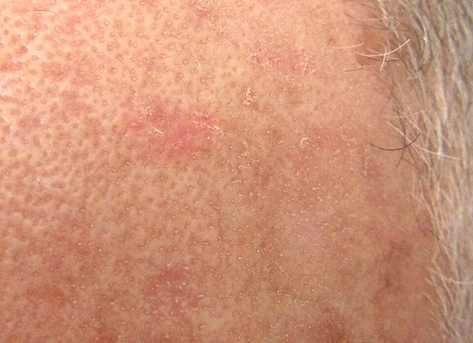
Actinic keratoses present as rough, scaly, erythematous or skin-coloured macules or thin papules, typically a few millimetres to around a centimetre, with a characteristic gritty, 'sandpaper-like' texture that is often easier to detect by running a fingertip over the skin than by looking at it. They occur on chronically sun-exposed sites - the face, scalp (especially in balding men), ears, dorsum of the hands and forearms.
Grading (Olsen scale)
Severity is graded using the Olsen scale, originally developed to standardise assessment of response in treatment trials.5
| Grade | Features |
|---|---|
| I (mild) | Barely palpable; more easily felt than seen |
| II (moderate) | Easily palpable and visible hyperkeratotic papule |
| III (severe/hyperkeratotic) | Thick, obviously visible hyperkeratosis, sometimes forming a cutaneous horn |
Variants
- Hypertrophic actinic keratosis - a markedly thickened, warty lesion that can be difficult to distinguish clinically from SCC, and often needs biopsy to be sure
- Pigmented actinic keratosis - brown pigmentation can cause confusion with a lentigo or early lentigo maligna
- Actinic cheilitis - the equivalent process affecting the lower lip, presenting as persistent dryness, scaling, and blurring of the normal sharp vermilion border; carries a comparatively higher risk of progression to SCC of the lip
- Cutaneous horn - a conical, keratotic projection that can arise from an actinic keratosis, a wart, or an underlying SCC, and always needs biopsy of its base to determine the underlying cause
Clinical examination
- Palpation as well as inspection - many actinic keratoses are more easily felt as a rough texture than seen, particularly early (grade I) lesions
- Distribution and number - single versus multiple lesions, and the overall extent of sun-damaged 'field' skin
- Induration, tenderness or ulceration - features that should prompt reconsideration of simple actinic keratosis in favour of possible SCC
- The lower lip - specifically examined for actinic cheilitis, which is easy to overlook
- Full skin examination for other sun-damage related lesions - solar lentigines, BCC, SCC and Bowen disease frequently coexist in the same chronically sun-exposed patient
Differential diagnosis
- Seborrhoeic keratosis - a 'stuck-on', warty surface with horn cysts on dermoscopy, generally thicker and more clearly demarcated
- Bowen disease - a larger, more persistent, well-demarcated erythematous scaly plaque, representing full-thickness in situ carcinoma rather than the more limited atypia of actinic keratosis
- Invasive squamous cell carcinoma - induration, tenderness, ulceration or rapid growth distinguish it from simple actinic keratosis
- Discoid lupus erythematosus - scaly, erythematous plaques with follicular plugging and scarring, usually in a younger patient and with other features of lupus
- Psoriasis - well-demarcated plaques with silvery scale, though the distribution and patient age usually differ from actinic keratosis
Investigations
Actinic keratosis is a clinical diagnosis in the great majority of cases. Biopsy is reserved for lesions where invasive SCC cannot be confidently excluded.
- Biopsy - indicated for any lesion that is indurated, tender, rapidly growing, ulcerated, unusually thick (hypertrophic), or fails to respond to appropriate treatment, since these features can indicate progression to invasive SCC
- Dermoscopy - can support the clinical diagnosis, typically showing a subtle pink-red pseudo-network with surrounding scale, though this is used more to support confidence in the diagnosis than to replace biopsy where there is genuine concern
Management
Treatment choice depends on the number, thickness and distribution of lesions, balancing efficacy against local skin reaction and cosmetic outcome.2
Lesion-directed treatment
Appropriate for a small number of discrete, well-defined lesions.
- Cryotherapy (liquid nitrogen) - quick, effective, and the most widely used lesion-directed treatment; can cause blistering, hypopigmentation or scarring, particularly with more aggressive freezing
- Curettage and cautery - useful for thicker (hypertrophic) lesions, and provides a tissue sample for histology, which cryotherapy does not
Field-directed treatment
Preferred where lesions are numerous, poorly defined, or field cancerisation is extensive, since it treats subclinical disease across the whole area rather than only the visible lesions.7
- Topical 5-fluorouracil - an antimetabolite applied over several weeks; produces a brisk inflammatory reaction (erythema, erosion) in treated areas, which is an expected part of an effective response rather than a complication, though it needs clear counselling in advance so patients are not alarmed6
- Topical imiquimod - an immune response modifier, applied over a similar timescale, also producing an inflammatory reaction as part of its mechanism
- Topical diclofenac gel - a gentler option with a milder inflammatory reaction, often better tolerated but generally less effective than 5-fluorouracil or imiquimod
- Photodynamic therapy - a topical photosensitiser activated with light, giving good cosmetic outcomes and useful for facial and scalp field disease, though it requires a hospital or clinic visit rather than being self-administered at home
General measures
- Sun protection - regular broad-spectrum sunscreen use, protective clothing and avoiding peak UV hours reduces the development of new lesions and is a central, ongoing part of management rather than an afterthought
- Periodic skin review - particularly for patients with multiple or high-grade lesions, immunosuppression, or a personal history of skin cancer
Complications
- Progression to invasive squamous cell carcinoma - the principal concern, though the annual risk per individual lesion is low
- Cosmetic and symptomatic impact - dryness, roughness and mild discomfort from the lesions themselves
- Local skin reactions to field-directed treatment - expected erythema, erosion and discomfort during a course of topical 5-fluorouracil or imiquimod, which can affect adherence if not explained clearly in advance
- Scarring or pigment change after cryotherapy, particularly with more aggressive freezing of thicker lesions
Red flags
Prognosis
The malignant transformation rate of an individual actinic keratosis to invasive SCC is low, classically quoted at well under 1% per lesion per year, and a meaningful proportion of lesions in fact regress spontaneously, particularly with improved sun protection.4
The practical significance of actinic keratosis is cumulative rather than dramatic: because lesions are so common and patients often have many over a lifetime, and because their presence marks a patient at genuinely higher overall risk of skin cancer of all types, actinic keratosis is best understood as a marker of a chronically sun-damaged 'field' at risk, rather than judged solely on the very low risk carried by any single lesion. With appropriate treatment and consistent sun protection, most patients do well, and the priority is long-term surveillance for new lesions and other skin cancers rather than anxiety about any one spot.
References
- NICE Clinical Knowledge Summaries. Actinic keratoses. Available here
- de Berker D, McGregor JM, Mohd Mustapa MF et al. British Association of Dermatologists' guidelines for the care of patients with actinic keratosis 2017. British Journal of Dermatology. 2017. Available here
- Berman B, Cockerell CJ. Pathobiology of actinic keratosis: clinical and histopathologic correlation. Journal of the American Academy of Dermatology. 2013. Available here
- Marks R, Rennie G, Selwood TS. Malignant transformation of solar keratoses to squamous cell carcinoma. The Lancet. 1988. Available here
- Olsen EA, Abernethy ML, Kulp-Shorten C et al. A double-blind, vehicle-controlled study evaluating masoprocol cream in the treatment of actinic keratoses. Journal of the American Academy of Dermatology. 1991. Available here
- BNF. Fluorouracil. Available here
- Gupta AK, Paquet M, Villanueva E, Brintnell W. Interventions for actinic keratoses. Cochrane Database of Systematic Reviews. 2012. Available here
- DermNet NZ. Actinic keratosis. Available here
This article is written for revision and education. It is not clinical guidance and must not be used to make decisions about the care of a patient. Always check current NICE guidance and local protocols.