
Right Heart Valve Disease: Tricuspid and Pulmonary Valves
Key points
- Right-sided murmurs: are louder on inspiration - RILE. Inspiration increases venous return to the right heart.
- Tricuspid regurgitation: by far the commonest right-sided lesion, and usually secondary to left heart disease or pulmonary hypertension rather than to the valve itself.
- The JVP tells you which: giant V waves mean tricuspid regurgitation; giant A waves mean tricuspid or pulmonary stenosis.
- Pulsatile hepatomegaly: is close to specific for severe tricuspid regurgitation.
- Intravenous drug use: causes right-sided endocarditis, typically Staphylococcus aureus on the tricuspid valve, with septic pulmonary emboli and often no murmur.
- Pulmonary stenosis: is nearly always congenital - isolated, or part of tetralogy of Fallot or Noonan syndrome. Treated with balloon valvuloplasty.
- Carcinoid heart disease: affects the right heart only, because serotonin is inactivated as blood passes through the lungs.
- Treat the cause: in secondary tricuspid regurgitation, managing the left heart and pulmonary hypertension comes before touching the valve.
Introduction
The tricuspid and pulmonary valves are neglected in teaching and in practice - tricuspid regurgitation has been called the forgotten valve lesion. They matter because right-sided disease is common, produces a characteristic and recognisable clinical picture, and usually points to a problem somewhere else.
The unifying rule for examination is RILE: Right-sided murmurs are louder on Inspiration, Left-sided on Expiration. Inspiration drops intrathoracic pressure and increases venous return to the right heart, so more blood crosses the right-sided valves and the murmur gets louder. For tricuspid regurgitation this is called Carvallo sign.
The second unifying rule is that most right-sided valve disease is secondary. Before diagnosing a tricuspid valve problem, ask what is raising the pressure or dilating the chamber behind it.
Reading the JVP
The jugular venous pressure is the most useful bedside tool in right heart disease, because the internal jugular vein is in direct continuity with the right atrium and therefore displays its pressure waveform.
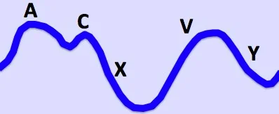
| Wave | Normally represents | Abnormality |
|---|---|---|
| a wave | Atrial contraction | Giant a waves: tricuspid stenosis, pulmonary stenosis, pulmonary hypertension. Absent: atrial fibrillation. Cannon a waves: complete heart block, when the atrium contracts against a closed tricuspid valve. |
| x descent | Atrial relaxation | Exaggerated in cardiac tamponade |
| v wave | Atrial filling against a closed tricuspid valve | Giant v waves: tricuspid regurgitation - blood is driven back into the atrium during systole |
| y descent | Atrial emptying as the tricuspid valve opens | Slow: tricuspid stenosis. Sharp: constrictive pericarditis. Absent: cardiac tamponade. |
Tricuspid regurgitation
The commonest right-sided lesion by a wide margin, and in most patients the valve itself is structurally normal.
| Secondary (functional) - the majority | Primary (valve disease) |
|---|---|
| Pulmonary hypertension of any cause | Infective endocarditis, classically in intravenous drug use |
| Left heart disease raising pulmonary pressures | Rheumatic heart disease - almost always with mitral disease |
| Right ventricular dilatation stretching the annulus | Carcinoid syndrome |
| Chronic atrial fibrillation with annular dilatation | Ebstein anomaly - a congenitally displaced valve |
| Right ventricular infarction | Pacemaker or ICD lead crossing the valve |
| Myxomatous degeneration; blunt chest trauma |
Clinical features
Symptoms are those of systemic venous congestion and low output: fatigue, ankle swelling, abdominal distension from ascites, right upper quadrant discomfort from hepatic congestion, nausea and early satiety. Breathlessness usually reflects the underlying left-sided or pulmonary cause rather than the tricuspid lesion itself.
- Raised JVP with giant v waves
- Pansystolic murmur at the lower left sternal edge, louder on inspiration (Carvallo sign)
- Right ventricular heave at the left sternal edge
- Pulsatile hepatomegaly
- Ascites and peripheral oedema, and in advanced cases jaundice and cardiac cirrhosis
- Atrial fibrillation is common
Management
For secondary regurgitation, treat what is driving it - optimise left heart failure therapy, treat pulmonary hypertension, control atrial fibrillation - and use loop diuretics for congestion. Improving the upstream problem often reduces the regurgitation without touching the valve.
Surgery is considered for severe symptomatic primary disease, and importantly is often performed at the same time as left-sided valve surgery, because operating on the tricuspid valve later as an isolated procedure carries a much higher mortality. Repair with annuloplasty is preferred to replacement. Transcatheter tricuspid repair and replacement are developing rapidly for patients unsuitable for surgery.
Tricuspid stenosis
Rare in isolation. Rheumatic heart disease is the commonest cause and virtually never affects the tricuspid valve alone - there is nearly always coexisting mitral disease, so finding tricuspid stenosis should prompt a careful search of the left heart. Other causes are carcinoid syndrome and, rarely, congenital disease.
- Raised JVP with giant a waves and a slow y descent
- Mid-diastolic murmur at the lower left sternal edge, louder on inspiration
- Hepatomegaly, ascites and peripheral oedema - often marked, and out of proportion to the breathlessness
- An opening snap may be audible
A useful clinical clue: a patient with rheumatic mitral stenosis whose lungs stay relatively clear while the abdomen and legs fill with fluid may have tricuspid stenosis limiting flow into the right ventricle, protecting the pulmonary circulation. Management is diuretics and, where severe, valve repair or replacement, usually combined with mitral surgery.
Pulmonary valve disease
Pulmonary stenosis
Almost always congenital. It occurs in isolation, or as part of a syndrome - and the associations are commonly examined:
- Tetralogy of Fallot - pulmonary stenosis, ventricular septal defect, overriding aorta and right ventricular hypertrophy
- Noonan syndrome - often with a dysplastic pulmonary valve
- Congenital rubella syndrome
- Williams syndrome - typically supravalvular
Acquired pulmonary stenosis is rare, seen with carcinoid syndrome and previously with rheumatic disease.
- Ejection systolic murmur in the 2nd left intercostal space, radiating to the left shoulder and back, louder on inspiration
- Ejection click, and widely split second heart sound with a soft P2 - right ventricular ejection takes longer
- Right ventricular heave and a giant a wave in the JVP
- Severe cases: fatigue, exertional breathlessness, syncope and right heart failure
Treatment of significant stenosis is balloon pulmonary valvuloplasty, which is highly effective and usually definitive - one of the more satisfying interventions in cardiology.
Pulmonary regurgitation
The commonest cause is pulmonary hypertension, which dilates the pulmonary annulus. The resulting high-pitched early diastolic murmur at the left sternal edge is the Graham Steell murmur, classically described in pulmonary hypertension secondary to mitral stenosis.
In adult practice the other major cause is previous repair of tetralogy of Fallot, where the surgery relieves the stenosis at the cost of a regurgitant valve. These patients need lifelong follow-up in an adult congenital heart disease service, since chronic regurgitation progressively dilates the right ventricle and eventually requires pulmonary valve replacement.
Mild pulmonary regurgitation is a common incidental echocardiographic finding in healthy people and needs no action.
Two causes worth knowing specifically
Right-sided infective endocarditis
Carcinoid heart disease
Neuroendocrine tumours with liver metastases release serotonin and other vasoactive mediators into the systemic venous circulation, causing fibrous plaque deposition on the right-sided endocardium. The result is tricuspid regurgitation and pulmonary stenosis, often together.
Investigations
- Echocardiography - the definitive test. Grades the lesion, estimates pulmonary artery systolic pressure from the tricuspid regurgitant jet, and assesses right ventricular size and function. Crucially it also examines the left heart, which is where the cause usually lies.
- ECG - right ventricular hypertrophy with right axis deviation and dominant R in V1; P pulmonale (tall peaked P waves) of right atrial enlargement; atrial fibrillation
- Chest X-ray - right heart enlargement; prominent pulmonary arteries in pulmonary hypertension; post-stenotic dilatation in pulmonary stenosis; cavitating nodules in septic emboli
- Bloods - FBC, U&Es, LFTs (congestive hepatopathy), NT-proBNP, and blood cultures where endocarditis is suspected. Urinary 5-HIAA if carcinoid is possible.
- Cardiac MRI - the reference standard for right ventricular volumes and function, and central to deciding when to replace a pulmonary valve after tetralogy repair
- Right heart catheterisation - to confirm and characterise pulmonary hypertension where that is the underlying question
Complications
- Right ventricular failure - progressive oedema, ascites and fatigue
- Congestive hepatopathy and cardiac cirrhosis - chronic hepatic venous congestion, with deranged LFTs and eventually irreversible fibrosis
- Atrial fibrillation and other arrhythmias - from right atrial dilatation
- Cardiac cachexia and malabsorption - from gut congestion
- Septic pulmonary emboli and lung abscess - in right-sided endocarditis
- Paradoxical embolism - where a patent foramen ovale allows right-to-left shunting as right-sided pressures rise
- Progressive right ventricular dilatation after tetralogy repair, which may become irreversible if valve replacement is left too late
Red flags
Prognosis
Prognosis in secondary tricuspid regurgitation is largely that of the underlying disease, but the regurgitation is not merely a marker - severe tricuspid regurgitation independently predicts worse survival even after adjusting for left ventricular function and pulmonary pressures.2 This has driven growing interest in treating it earlier rather than accepting it as an inevitable accompaniment.
Isolated late tricuspid surgery, performed once the right ventricle and liver are already failing, carries a high operative mortality. This is the argument for addressing significant tricuspid regurgitation at the time of left-sided valve surgery rather than returning to it years later.
Congenital pulmonary stenosis has an excellent outlook: balloon valvuloplasty in childhood or adulthood is usually definitive, and long-term survival is close to normal. Right-sided endocarditis in intravenous drug use generally responds better to antibiotics than left-sided disease and requires surgery less often, but the prognosis is dominated by whether the underlying drug use can be addressed.
References
- Vahanian A, Beyersdorf F, Praz F et al. ESC/EACTS Guidelines for the management of valvular heart disease. European Heart Journal. 2021. Available here
- Nath J, Foster E, Heidenreich PA. Impact of tricuspid regurgitation on long-term survival. Journal of the American College of Cardiology. 2004. Available here
- NICE NG208. Heart valve disease presenting in adults: investigation and management. 2021. Available here
- Delgado V, Ajmone Marsan N, de Waha S et al. ESC Guidelines for the management of endocarditis. European Heart Journal. 2023. Available here
- Baumgartner H, De Backer J, Babu-Narayan SV et al. ESC Guidelines for the management of adult congenital heart disease. European Heart Journal. 2021. Available here
- NICE Clinical Knowledge Summaries. Heart murmurs. Available here
- Davar J, Connolly HM, Caplin ME et al. Diagnosing and managing carcinoid heart disease. Journal of the American College of Cardiology. 2017. Available here
This article is written for revision and education. It is not clinical guidance and must not be used to make decisions about the care of a patient. Always check current NICE guidance and local protocols.