
Atrial Flutter: Recognition and Management
Key points
- Atrial flutter: an organised macro-re-entrant atrial tachycardia, usually circling the tricuspid annulus in the right atrium at about 300 beats per minute.
- ECG: sawtooth flutter waves, best seen in the inferior leads (II, III, aVF) and V1, with no isoelectric baseline between them.
- The 150 rule: a regular narrow complex tachycardia at almost exactly 150 beats per minute is atrial flutter with 2:1 block until proven otherwise.
- Unmasking it: vagal manoeuvres or adenosine transiently increase AV block and reveal the flutter waves. Adenosine is diagnostic here, not therapeutic.
- Typical flutter: counterclockwise re-entry gives negative sawtooth waves in the inferior leads and positive flutter waves in V1.
- Stroke risk: the same as atrial fibrillation. Assess with CHA2DS2-VASc and anticoagulate on the same criteria - this is a frequently missed point.
- Acute management: identical principles to AF - cardiovert if unstable, and observe the 48-hour rule before elective cardioversion. Flutter cardioverts at lower energies than AF.
- Definitive treatment: cavotricuspid isthmus ablation cures typical flutter in over 90% of cases and is offered early, because rate control is notoriously difficult.
Introduction
Atrial flutter is an organised atrial tachyarrhythmia caused by a macro-re-entrant circuit within the atria. Unlike atrial fibrillation, where atrial activity is chaotic and disorganised, in flutter the atria depolarise in a regular, coordinated wave that travels around a fixed anatomical circuit at roughly 300 beats per minute.
That distinction - organised versus chaotic - explains almost every difference between the two conditions. Flutter produces regular, uniform atrial waves on the ECG rather than a chaotic baseline; it responds to lower cardioversion energies; and because the circuit is anatomically defined, it can be interrupted permanently by a single line of ablation.
Flutter and fibrillation frequently coexist in the same patient, and one often degenerates into the other. This means that curing flutter by ablation does not guarantee freedom from AF, and it is one reason anticoagulation decisions are made on the same basis for both.
Aetiology and mechanism
The circuit
Typical (type I) atrial flutter accounts for around 90% of cases. The re-entrant wavefront circles the tricuspid annulus in the right atrium, passing through a narrow strip of tissue between the tricuspid valve and the inferior vena cava called the cavotricuspid isthmus. This isthmus is the vulnerable point of the circuit and the target of ablation.
- Counterclockwise typical flutter (the commoner form) - the wavefront travels up the interatrial septum and down the right atrial free wall, producing negative sawtooth waves in the inferior leads and positive flutter waves in V1
- Clockwise typical flutter - the reverse direction, producing positive flutter waves in the inferior leads and negative waves in V1
Atypical flutter uses a circuit elsewhere, often in the left atrium or around scar from previous cardiac surgery or a prior AF ablation. It does not involve the cavotricuspid isthmus, so isthmus ablation does not cure it, and the ECG appearance is more variable.
Causes and associations
The risk factors are essentially those for atrial fibrillation, since both reflect an abnormal atrial substrate.
- Ischaemic heart disease and previous myocardial infarction
- Hypertension with left atrial enlargement
- Heart failure and cardiomyopathy
- Valvular heart disease, particularly mitral and tricuspid disease
- Chronic lung disease and cor pulmonale - right atrial dilatation is a strong association, given the right atrial circuit
- Pulmonary embolism
- Thyrotoxicosis
- Alcohol excess
- Post-cardiac surgery - flutter around surgical scar (incisional flutter) is common after congenital heart surgery
- After AF ablation - iatrogenic left atrial flutter around ablation lines
- Obesity and obstructive sleep apnoea
Clinical features
Symptoms depend almost entirely on the ventricular rate and the state of the underlying heart. Flutter with 4:1 block at 75/min may be entirely asymptomatic; flutter with 2:1 block at 150/min in a patient with impaired ventricular function may precipitate pulmonary oedema.
Symptoms
- Palpitations - typically described as regular and racing, in contrast to the irregular sensation of AF
- Breathlessness and reduced exercise tolerance
- Fatigue
- Chest pain - from increased myocardial oxygen demand
- Presyncope or syncope - uncommon but possible, particularly if 1:1 conduction occurs
- Asymptomatic - found incidentally, particularly with a high degree of AV block
Examination
- A regular tachycardia, classically at 150/min. Note that with variable AV block the pulse is irregular and clinically indistinguishable from AF.
- Flutter waves in the JVP - rapid, regular a waves at around 300/min may be visible as a fine flicker in the neck. This is an uncommon but striking sign.
- Signs of the underlying cause - chronic lung disease, heart failure, valve disease, thyrotoxicosis
- Signs of decompensation - hypotension, pulmonary oedema, poor peripheral perfusion
Investigations
ECG
The 12-lead ECG is diagnostic when the flutter waves are visible.
- Sawtooth flutter waves (F waves) at approximately 300/min, best seen in leads II, III and aVF and in V1
- No isoelectric baseline between the flutter waves in the inferior leads - the atrial activity is continuous. This is the feature that distinguishes flutter from atrial tachycardia, where discrete P waves are separated by a flat baseline.
- Regular ventricular response if the AV block ratio is fixed (2:1, 3:1 or 4:1), and irregular if it varies
- Narrow QRS complexes, unless there is bundle branch block or aberrant conduction
- Even-numbered conduction ratios are more common than odd, so 2:1 and 4:1 predominate
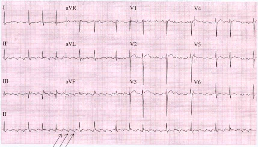
Unmasking flutter waves
If the diagnosis is uncertain, increasing the degree of AV block slows the ventricular rate transiently and exposes the underlying atrial activity.
- Vagal manoeuvres - the modified Valsalva manoeuvre or carotid sinus massage (avoiding the latter if there is a carotid bruit or previous stroke or TIA)
- Adenosine 6 mg IV as a rapid bolus with a saline flush, escalating to 12 mg if needed
Further investigations
- U&Es, magnesium, FBC and thyroid function - as for atrial fibrillation
- Transthoracic echocardiogram - to assess atrial size, ventricular function and valve disease
- Transoesophageal echocardiogram - to exclude atrial thrombus if early cardioversion is planned in flutter of over 48 hours or unknown duration
- Chest X-ray - looking for chronic lung disease, heart failure or another precipitant
- Ambulatory monitoring - for paroxysmal flutter, and to assess rate control
- Consider CTPA - flutter with hypoxia and breathlessness may be the presentation of a pulmonary embolism
Differential diagnosis
| Rhythm | Atrial rate | Atrial activity | Ventricular rhythm |
|---|---|---|---|
| Atrial flutter | ~300/min | Continuous sawtooth, no isoelectric baseline | Regular if fixed block; irregular if variable |
| Atrial fibrillation | 300-600/min | Chaotic fibrillatory baseline, no discrete waves | Irregularly irregular |
| Sinus tachycardia | 100-160/min | Normal upright P waves in II with an isoelectric baseline | Regular; rate varies with the clinical state |
| Atrial tachycardia | 150-250/min | Discrete abnormal P waves with an isoelectric baseline between | Regular or variable |
| AVNRT | 140-250/min | P waves hidden in the QRS, or a pseudo-R in V1 | Regular; abrupt onset and offset |
| Multifocal atrial tachycardia | 100-150/min | Three or more distinct P wave morphologies | Irregular; classically in severe COPD |
Management
Acute management follows the same structure as atrial fibrillation, with two practical differences: flutter is considerably harder to rate control with drugs, and it responds to lower cardioversion energies.
The unstable patient
Rate control
Use a beta-blocker or a rate-limiting calcium channel blocker (diltiazem or verapamil), as in AF. In practice, rate control in flutter is frequently unsatisfactory: because conduction occurs in discrete ratios, partially blocking the AV node often shifts the patient abruptly from 2:1 to 3:1 conduction rather than producing a smooth reduction in rate. This difficulty is a major reason why definitive treatment is pursued earlier in flutter than in AF.
Rhythm control
- Synchronised DC cardioversion - highly effective, and the preferred method
- Overdrive atrial pacing - possible if the patient already has a pacemaker or temporary atrial wires after cardiac surgery
- Pharmacological cardioversion - less reliable than in AF. Amiodarone may be used; flecainide should be given only with an AV nodal blocking agent, because it slows the atrial rate and can permit 1:1 conduction with a catastrophic rise in ventricular rate.
- The 48-hour rule applies exactly as in AF - anticoagulate for at least 3 weeks before elective cardioversion, or exclude thrombus with a transoesophageal echocardiogram, and continue anticoagulation for at least 4 weeks afterwards
Anticoagulation
Stroke risk in atrial flutter is regarded as equivalent to that in atrial fibrillation, and anticoagulation is assessed and prescribed on exactly the same basis: CHA2DS2-VASc, with a DOAC first line unless there is moderate to severe mitral stenosis or a mechanical valve, in which case warfarin is used.2
This point is frequently missed in practice. The mechanical activity of the atria in flutter is more organised than in AF but remains grossly impaired, and many patients with flutter also have episodes of AF, whether documented or not.
Catheter ablation
Cavotricuspid isthmus ablation is the definitive treatment for typical atrial flutter. A line of radiofrequency lesions across the isthmus creates bidirectional conduction block, making the circuit impossible to sustain.
- Success rates exceed 90%, considerably higher than for AF ablation, because the target is a single well-defined anatomical structure
- It is offered early - often after a first symptomatic episode - given the difficulty of drug rate control and the low complication rate
- Anticoagulation continues after ablation if the CHA2DS2-VASc score warrants it. Up to a third of patients develop atrial fibrillation in the years after successful flutter ablation, since the underlying atrial substrate is unchanged.
- Atypical flutter requires more complex mapping and ablation with lower success rates
Treat the underlying cause
Correct thyrotoxicosis, optimise treatment of chronic lung disease and heart failure, address alcohol excess, obesity and obstructive sleep apnoea, and reassess after any acute precipitant such as sepsis or surgery has resolved. Flutter that occurs solely in the context of an acute illness may not recur.
Complications
- Ischaemic stroke and systemic embolism - equivalent risk to atrial fibrillation
- Tachycardia-induced cardiomyopathy - particularly relevant in flutter, because a persistent 2:1 conduction ratio can maintain a rate of 150/min for weeks while causing relatively modest symptoms. The resulting dilated cardiomyopathy is largely reversible once rhythm or rate is controlled.
- Heart failure decompensation
- 1:1 conduction - a life-threatening complication, usually iatrogenic after class Ic antiarrhythmic therapy, and occasionally seen with high sympathetic drive or an accessory pathway
- Progression to atrial fibrillation - very common, and a reason for continued follow-up after ablation
- Myocardial ischaemia in patients with coronary disease
- Complications of ablation - vascular access complications, cardiac tamponade, and the small risk of complete heart block
Red flags
Prognosis
The prognosis of atrial flutter is largely that of the underlying heart disease. In an otherwise healthy patient, isthmus ablation is curative in over 90% of cases with a very low complication rate, and most patients return to a normal quality of life without antiarrhythmic drugs.
The two caveats are important. First, atrial fibrillation develops in a substantial minority after successful flutter ablation - reported in up to 30% over several years - because ablation removes the flutter circuit but does not change the diseased atrial substrate that produced it. Second, anticoagulation decisions are not altered by a successful ablation: they continue to be driven by CHA2DS2-VASc.
Untreated flutter with a persistently rapid ventricular response carries a real risk of tachycardia-induced cardiomyopathy, which is why a rate that seems tolerable to the patient should not be accepted indefinitely without an echocardiogram.
References
- Resuscitation Council UK. Adult advanced life support guidelines: peri-arrest arrhythmias. Available here
- NICE NG196. Atrial fibrillation: diagnosis and management. 2021, updated 2023. Available here
- Brugada J, Katritsis DG, Arbelo E et al. 2019 ESC Guidelines for the management of patients with supraventricular tachycardia. European Heart Journal. 2020. Available here
- Hindricks G, Potpara T, Dagres N et al. 2020 ESC Guidelines for the diagnosis and management of atrial fibrillation. European Heart Journal. 2021. Available here
- BNF. Flecainide acetate - indications and cautions. Available here
- BNF. Adenosine - indications and cautions. Available here
- NICE Clinical Knowledge Summaries. Atrial fibrillation. Available here
- DVLA. Assessing fitness to drive: a guide for medical professionals. Available here
This article is written for revision and education. It is not clinical guidance and must not be used to make decisions about the care of a patient. Always check current NICE guidance and local protocols.