
Gastrointestinal Perforation
Key points
- Perforation: a full-thickness breach of the gastrointestinal wall, releasing luminal contents into the peritoneal or mediastinal cavity.
- Commonest causes: perforated peptic ulcer, diverticular disease, appendicitis, obstructing or perforating colorectal cancer, and iatrogenic injury at endoscopy.
- Classic presentation: sudden, severe, generalised abdominal pain in a patient who lies still, with a board-like rigid abdomen and shock.
- Two-phase peritonitis: an initial chemical peritonitis from acid or bile, followed within hours by bacterial peritonitis as enteric organisms multiply.
- Erect chest radiograph: shows free subdiaphragmatic gas in only about 70 to 80% of cases, so a normal film never excludes perforation.
- Definitive imaging: contrast-enhanced CT of the abdomen and pelvis, which detects even tiny volumes of free gas and localises the site.
- Management: aggressive resuscitation, broad-spectrum antibiotics, analgesia, nasogastric decompression and urgent surgery for source control.
- Peptic ulcer repair: an omental (Graham) patch closes the defect, followed by peritoneal lavage, Helicobacter pylori eradication and a proton pump inhibitor.
Introduction
Gastrointestinal perforation is a full-thickness breach of the wall of a hollow viscus, allowing gas, acid, bile, enzymes and enteric bacteria to escape into a cavity that is normally sterile. It is one of the few genuinely time-critical diagnoses on the surgical take, and the mortality curve is steep: outcome deteriorates measurably with each hour of delay to source control.
The presentation is often described as one of the most striking in medicine - a patient who can name the minute the pain began, who lies rigidly still, and whose abdomen feels like a board. In practice that textbook picture is common in younger patients with a perforated duodenal ulcer and much less common in the older, frailer or immunosuppressed patients who make up a large share of cases.
The distinction that governs everything is between free perforation, which spills into the general peritoneal cavity and produces generalised peritonitis, and contained or sealed perforation, where omentum or adjacent viscera wall off the leak, producing a localised abscess or phlegmon. The first almost always needs an operation; the second may not.
Causes
| Site | Causes |
|---|---|
| Oesophagus | Iatrogenic at endoscopy or dilatation (the commonest cause), Boerhaave syndrome from forceful vomiting, foreign body or caustic ingestion, malignancy |
| Stomach and duodenum | Peptic ulcer disease, NSAIDs and corticosteroids, Helicobacter pylori, gastric carcinoma, iatrogenic at endoscopy, trauma |
| Small bowel | Trauma, strangulated hernia, Crohn disease, mesenteric ischaemia, closed loop obstruction, foreign body, typhoid and tuberculosis worldwide |
| Appendix | Untreated acute appendicitis |
| Colon | Diverticular disease, colorectal carcinoma, closed loop obstruction with caecal blowout, volvulus, colonoscopic injury, severe colitis with toxic megacolon, stercoral perforation from impacted faeces |
| Any site | Blunt or penetrating trauma, ischaemia, radiation enteritis, ingested foreign body |
Two causes deserve particular attention because they are common and often missed. Perforated peptic ulcer remains frequent despite effective acid suppression, largely because of NSAID use, and the anterior wall of the first part of the duodenum is the classic site since it has no retroperitoneal structure behind it to seal a leak. Iatrogenic colonic perforation at colonoscopy occurs in roughly one in a thousand diagnostic procedures and more often after polypectomy, and should be considered in anyone with abdominal pain after endoscopy.
Pathophysiology
The consequences of a perforation depend on what escapes and where it goes, and this proceeds in two overlapping phases.
- Chemical peritonitis, in the first few hours. Gastric acid, bile and pancreatic enzymes are intensely irritant to the parietal peritoneum, producing immediate severe pain, reflex rigidity of the abdominal wall and a large exudative fluid shift into the peritoneal cavity. The patient may become profoundly hypovolaemic before any infection has developed.
- Bacterial peritonitis, from around 6 to 12 hours. Enteric organisms proliferate in the peritoneal exudate. The bacterial load rises with distance down the gut, so a duodenal perforation contaminates with relatively few organisms while a colonic perforation delivers a massive faecal inoculum, which is why colonic perforation carries far higher mortality.
- Systemic inflammatory response and septic shock, with vasodilatation, capillary leak, myocardial depression, acute kidney injury and multi-organ failure
- Paralytic ileus, which compounds the fluid loss and vomiting
Occasionally there is a deceptive quiet phase a few hours after the initial event, when the chemical irritation has been diluted by peritoneal exudate and the pain temporarily eases. Patients and inexperienced clinicians can be reassured by this, only for the picture to deteriorate sharply as bacterial peritonitis establishes. Never accept improvement alone as evidence that a perforation has sealed.
Clinical features
History
- Sudden onset pain, maximal within seconds and often precisely timed by the patient. This instantaneous onset is the hallmark.
- Initially localised, rapidly becoming generalised as contents spread through the peritoneal cavity
- Worse on any movement, so the patient lies still and dislikes coughing or the journey to hospital
- Shoulder tip pain from diaphragmatic irritation of the phrenic nerve
- Nausea and vomiting, and later absolute constipation from the accompanying ileus
- Preceding symptoms - dyspepsia and NSAID use before a peptic ulcer perforation, left iliac fossa pain and altered bowel habit before a diverticular one, forceful vomiting before Boerhaave syndrome, or a recent endoscopy
Examination
- The patient lies motionless, often with shallow breathing because deep inspiration moves the diaphragm against inflamed peritoneum
- Signs of shock - tachycardia, hypotension, cool peripheries, oliguria and a raised NEWS2 score
- A rigid, board-like abdomen with generalised guarding, involuntary and not overcome by distraction
- Percussion tenderness throughout and absent bowel sounds
- Loss of liver dullness to percussion in the right upper quadrant, where free gas has interposed between the liver and the abdominal wall. This is a specific but insensitive sign.
- Subcutaneous emphysema in the neck and a mediastinal crunch on auscultation (Hamman sign) in oesophageal perforation
- Pyrexia, though older and immunosuppressed patients may be normothermic or hypothermic
Investigations
Investigation must not delay resuscitation, and in a patient with generalised peritonitis and shock the decision to operate can be made clinically.
Blood tests
- FBC - leucocytosis, though a low or normal white cell count in a very unwell patient is an ominous sign of overwhelming sepsis
- U&Es - acute kidney injury from hypovolaemia and sepsis
- Venous or arterial blood gas - lactate and base deficit are the best early markers of hypoperfusion and guide the urgency of resuscitation
- Amylase or lipase - a moderately raised amylase occurs in perforation and can be mistaken for pancreatitis. A level more than three times normal usually indicates pancreatitis, but the two can coexist and CT settles the question.
- CRP, LFTs, clotting, group and crossmatch, and blood cultures before antibiotics
Imaging
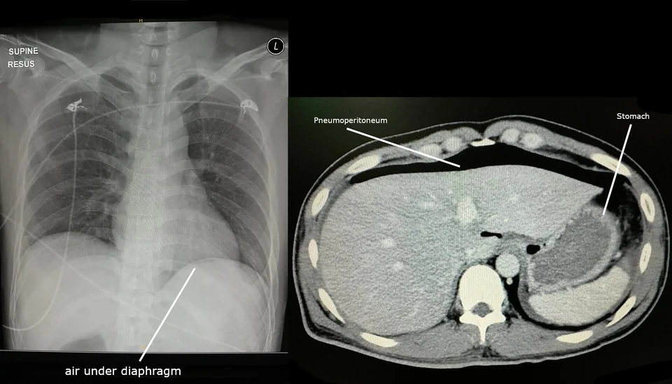
- Erect chest radiograph - quick and available anywhere, showing a crescent of free gas beneath the diaphragm. Its sensitivity is only around 70 to 80%, and it is lower still for small-bowel and retroperitoneal perforations, so a normal film does not exclude perforation. A left lateral decubitus film is an alternative in a patient who cannot sit up.
- Abdominal radiograph - may show Rigler sign, gas outlining both the mucosal and serosal surfaces of the bowel wall, or the falciform ligament sign, but it is not the test of choice
- Contrast-enhanced CT of the abdomen and pelvis is the definitive investigation. It detects volumes of free gas far too small to see on plain films, localises the perforation, distinguishes free from contained leaks, identifies an underlying cause such as a tumour or diverticular disease, and guides whether radiological drainage is feasible.1
- CT with water-soluble oral or rectal contrast where the site of a leak needs to be demonstrated, such as after anastomotic surgery or suspected iatrogenic colonic injury
- CT of the chest with contrast swallow for suspected oesophageal perforation, which may show mediastinal gas, pleural effusion and contrast extravasation
Management
Immediate resuscitation
- A to E assessment, oxygen, continuous monitoring and early critical care involvement
- Two large-bore cannulae and aggressive intravenous crystalloid resuscitation, guided by lactate clearance and urine output rather than by a fixed volume
- Broad-spectrum intravenous antibiotics within one hour, covering Gram-negative and anaerobic organisms according to local policy, after blood cultures2
- Nil by mouth with a nasogastric tube on free drainage, which reduces ongoing contamination and the risk of aspiration at induction
- Intravenous proton pump inhibitor where a peptic ulcer is the suspected source
- Titrated intravenous opioid analgesia and an antiemetic
- Urinary catheter and hourly urine output measurement
- Correct coagulopathy, discuss anticoagulant reversal, and send a group and crossmatch
- Urgent senior surgical review and a documented risk assessment before theatre
Source control
The principle of surgery is source control: close or resect the defect, remove contamination by peritoneal lavage, and drain any residual collection. The specific operation depends on the site.
| Site | Usual operation | Notes |
|---|---|---|
| Perforated duodenal ulcer | Omental (Graham) patch closure and peritoneal lavage, laparoscopic or open | Biopsy is not required. Helicobacter pylori eradication and a proton pump inhibitor afterwards, with repeat testing to confirm clearance. |
| Perforated gastric ulcer | Patch repair with biopsy of the ulcer edge, or local excision | Always biopsy - a perforated gastric ulcer may be a malignancy |
| Perforated appendix | Appendicectomy with lavage | Post-operative antibiotics for three to five days in complicated disease |
| Perforated diverticular disease | Laparoscopic lavage for purulent peritonitis in selected cases, or Hartmann procedure for faecal peritonitis | Faecal peritonitis with an unstable patient means resection, not lavage |
| Perforated colorectal cancer | Resection of the segment with an oncological margin, usually with a stoma | An emergency oncological resection carries a worse long-term outcome than an elective one |
| Small bowel perforation | Primary repair or segmental resection with anastomosis or stoma | Depends on contamination, bowel viability and physiological state |
| Oesophageal perforation | Primary repair with drainage, endoscopic stenting, or conservative management of a contained leak | Needs upper gastrointestinal or thoracic surgical input and intensive care |
Non-operative management
Selected patients can be managed without surgery, and the criteria are strict. This is reasonable where the perforation is contained and sealed on CT, the patient is haemodynamically stable without generalised peritonitis, and there is a facility for close monitoring with serial examination and repeat imaging. It applies most often to sealed peptic ulcer perforations, contained diverticular perforations with a small abscess amenable to percutaneous drainage, and some iatrogenic perforations recognised and clipped at the time of endoscopy.
Non-operative management is also chosen for palliative reasons in patients too frail or too unwell for surgery, or where an operation is not in keeping with the patient's wishes or ceiling of care. That decision should be made by a senior clinician, discussed openly with the patient and family, and accompanied by a clear plan for symptom control.
Complications
- Septic shock and multi-organ failure, the commonest cause of death
- Intra-abdominal or pelvic abscess, presenting several days later with swinging fever and ileus and usually treated by radiological drainage
- Acute kidney injury and acute respiratory distress syndrome as part of the systemic response
- Anastomotic leak and repair breakdown, particularly where bowel was oedematous or the patient malnourished
- Wound infection, wound dehiscence and incisional hernia, all common after emergency laparotomy for contamination
- Prolonged ileus and the need for parenteral nutrition
- Enterocutaneous fistula, a difficult and protracted complication
- Mediastinitis, empyema and oesophagopleural fistula after oesophageal perforation
- Adhesional obstruction and stoma complications in the longer term
Red flags
Prognosis
Prognosis is determined by three factors: how contaminated the peritoneum is, how physiologically robust the patient is, and how long the delay to source control has been. A young patient with a perforated duodenal ulcer repaired within a few hours generally does well. An older patient with faecal peritonitis from a perforated sigmoid carcinoma, several comorbidities and a delay of a day has a mortality that may exceed 30%.
For perforated peptic ulcer specifically, the Boey score is a simple and widely used tool that assigns one point each for shock on admission, major medical comorbidity, and duration of symptoms over 24 hours. Mortality rises steeply with each point, from a few per cent at zero to well over 50% at three, and it is a useful shorthand for why delay matters so much.3
The recurring theme, as with emergency laparotomy generally, is that the modifiable determinants of survival are organisational rather than technical. Recognising the diagnosis, resuscitating properly, giving antibiotics within the hour, getting the patient to CT and then to theatre without drift, and involving critical care early are all achievable on any surgical take, and together they matter more than the choice between one operation and another.
References
- Royal College of Radiologists. iRefer: making the best use of clinical radiology. Available here
- Sartelli M, Coccolini F, Kluger Y et al. WSES/GAIS/SIS-E/WSIS/AAST global clinical pathways for patients with intra-abdominal infections. World Journal of Emergency Surgery. 2021. Available here
- Tarasconi A, Coccolini F, Biffl WL et al. Perforated and bleeding peptic ulcer: WSES guidelines. World Journal of Emergency Surgery. 2020. Available here
This article is written for revision and education. It is not clinical guidance and must not be used to make decisions about the care of a patient. Always check current NICE guidance and local protocols.