
Gangrene and Necrotising Soft Tissue Infection
Key points
- Gangrene: death of tissue caused by a critical loss of blood supply, by overwhelming infection, or by both together.
- Dry gangrene: pure ischaemic necrosis with no infection. The tissue mummifies, blackens and demarcates sharply. There is time to plan.
- Wet gangrene: necrosis with superimposed bacterial infection. Swollen, blistered, malodorous and spreading, with systemic sepsis. There is no time.
- Gas gangrene: clostridial myonecrosis, usually Clostridium perfringens, whose alpha toxin destroys muscle. Crepitus, a brown discharge and rapid progression.
- Necrotising fasciitis: infection tracking along fascial planes. Type I is polymicrobial, type II is group A streptococcal. Pain out of proportion is the earliest sign.
- The diagnostic rule: necrotising infection is a clinical and surgical diagnosis. Normal-looking overlying skin does not exclude it, and imaging must never delay theatre.
- Treatment: for wet, gas or necrotising disease: resuscitation, broad-spectrum antibiotics with clindamycin for toxin suppression, and radical surgical debridement.
- Dry gangrene management: revascularise if possible, then amputate at a viable level or allow auto-amputation. Antibiotics are not required unless infection supervenes.
Introduction and classification
Gangrene is death of macroscopic tissue, and the clinically useful classification is by whether infection is present, because that single question determines whether the patient can be planned for an elective operation next week or needs to be in theatre within the hour.
| Type | Mechanism | Appearance | Urgency |
|---|---|---|---|
| Dry | Ischaemic necrosis without infection, usually from chronic arterial occlusion | Black, dry, shrunken, mummified tissue with a clear line of demarcation from viable skin. Not swollen and not malodorous. | Semi-elective - revascularise and plan amputation |
| Wet | Ischaemic or injured tissue colonised by bacteria, often in the presence of venous congestion or diabetes | Swollen, boggy, blistered, discoloured and foul-smelling, with poorly defined margins and surrounding cellulitis | Emergency - urgent debridement or amputation |
| Gas | Clostridial myonecrosis, most often Clostridium perfringens, following a contaminated wound or a bowel source | Tense, bronze or purple skin, tense blisters, a thin brown watery discharge with a sweetish odour, and crepitus | Extreme emergency - immediate radical debridement |
| Necrotising fasciitis | Rapidly spreading infection along fascial planes with thrombosis of perforating vessels | Pain far out of proportion to appearance, initially unremarkable skin, then dusky discolouration, bullae and anaesthesia | Extreme emergency - immediate surgical exploration |
| Internal (visceral) | Ischaemic necrosis of a viscus - bowel, gallbladder, testis | Not visible. Presents as an acute abdomen or acute scrotum with sepsis. | Emergency laparotomy or exploration |
Fournier gangrene is necrotising fasciitis of the perineum, scrotum and perianal region, and deserves separate mention because it is frequently the presentation of an unrecognised anorectal or urological source in a diabetic patient.
Aetiology and risk factors
Causes of ischaemic necrosis
- Peripheral arterial disease with chronic limb-threatening ischaemia - the commonest cause of dry gangrene in UK practice
- Acute limb ischaemia from embolus or thrombosis
- Diabetes mellitus, through the combination of distal arterial disease, neuropathy that hides injury, and impaired immunity
- Vasculitis, scleroderma and Raynaud phenomenon with critical digital ischaemia
- Frostbite and burns
- Ergotism, and vasopressor-induced peripheral ischaemia in the critically ill
- Compartment syndrome and crush injury
- Meningococcal septicaemia with purpura fulminans, particularly in children
Factors predisposing to infective necrosis
- Diabetes mellitus - the single most consistent association across necrotising infections and Fournier gangrene
- Immunosuppression - corticosteroids, chemotherapy, HIV, malignancy, neutropenia
- Intravenous drug use, particularly with skin popping or contaminated injection
- Trauma and surgery, especially with contaminated or devitalised tissue
- Obesity, alcohol excess, malnutrition and chronic kidney disease
- Peripheral vascular disease and chronic venous ulceration
- Recent varicella infection or NSAID use in children - a recognised association with group A streptococcal necrotising fasciitis
Clinical features
Dry gangrene
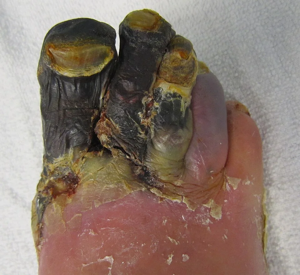
- Progression from pallor to dusky blue to black, over days or weeks
- Dry, shrivelled, mummified tissue which is cold and hard
- A sharp line of demarcation between dead and living tissue, which becomes clearer over time
- Preceding rest pain, which often diminishes once the nerves have died
- Absent pulses and other signs of chronic ischaemia - hair loss, shiny skin, thickened nails
- No swelling, no crepitus, no discharge and no systemic upset
Wet gangrene
- Swelling, blistering and a boggy texture, with skin that is discoloured rather than uniformly black
- Foul, putrid discharge and a distinctive smell
- Poorly demarcated, spreading erythema with surrounding cellulitis and lymphangitis
- Systemic sepsis - fever, tachycardia, hypotension and confusion
- Rapid progression over hours, in contrast to the indolent course of dry gangrene
Necrotising fasciitis and gas gangrene
In gas gangrene specifically, the incubation period after a contaminated wound is short - often less than 24 hours. The affected muscle is initially pale and non-contractile, later becoming brick red and then black, and does not bleed when cut. The classic thin, brown, sweet-smelling watery discharge, sometimes described as dishwater fluid, is distinct from the frank pus of an abscess.
Investigations
In suspected necrotising infection, investigations run alongside resuscitation and must never delay surgical exploration.
- FBC - marked leucocytosis, or leucopenia in overwhelming sepsis; anaemia and thrombocytopenia
- U&Es, creatinine, glucose and HbA1c - acute kidney injury is common, and undiagnosed diabetes is frequently found
- CRP - usually very high in necrotising infection
- Creatine kinase - raised in myonecrosis and a useful marker of muscle involvement
- Venous or arterial gas with lactate, and a clotting screen
- Blood cultures and deep tissue cultures taken at operation. Superficial swabs are unhelpful and may mislead.
- Plain radiograph - may show soft tissue gas, but is normal in most cases of necrotising fasciitis
- CT or MRI - shows fascial thickening, fluid tracking along fascial planes and gas. Useful only in the patient who is stable and where the diagnosis is genuinely uncertain; never as a reason to delay theatre.
- ABPI, duplex ultrasound and CT angiography where the underlying problem is arterial, to assess whether revascularisation is possible before deciding an amputation level
Management
Dry gangrene
The tissue is already dead, so the questions are whether the perfusion of the remaining limb can be improved and where to draw the line.
- Assess and treat the arterial supply. Urgent vascular referral with duplex and angiography, and revascularisation by angioplasty or bypass wherever feasible, since a better inflow may allow a more distal amputation or none at all.
- Analgesia, which is often substantial and may need neuropathic agents
- Meticulous wound care and offloading, keeping the tissue dry and protected, with specialist podiatry and tissue viability involvement
- Antibiotics are not indicated for uninfected dry gangrene, and reflex prescribing simply selects resistant organisms. Treat promptly if it becomes wet.
- Amputation at a viable level once demarcation is established and perfusion is optimised. Auto-amputation, in which a mummified digit separates spontaneously, is an acceptable strategy for a single toe in a patient unfit for surgery.
- Secondary prevention - smoking cessation, statin, antiplatelet, glycaemic control - and referral to the multidisciplinary diabetic foot service with a documented foot protection plan3
- Rehabilitation planning from the outset where a major amputation is likely, involving physiotherapy, occupational therapy and the prosthetics service
Wet gangrene, gas gangrene and necrotising fasciitis
These are managed identically in principle, because the principle is source control. Antibiotics cannot penetrate tissue whose blood supply has thrombosed, which is precisely what has happened in the affected fascia and muscle, so surgery is the treatment and antibiotics are the adjunct.2
- A to E assessment with aggressive fluid resuscitation, and immediate critical care involvement. These patients need substantial volumes and often vasopressor support.
- Blood cultures, then broad-spectrum intravenous antibiotics within the hour - typically a carbapenem or piperacillin-tazobactam plus clindamycin, following local microbiology policy and with early microbiology advice
- Clindamycin is given specifically for its antitoxin effect, suppressing streptococcal and clostridial exotoxin production by inhibiting protein synthesis. It works even against organisms in stationary phase, when beta-lactams are least effective.
- Immediate radical surgical debridement, removing all necrotic tissue back to healthy bleeding tissue. This is the intervention that determines survival, and delay of even a few hours measurably increases mortality.
- Mark the extent of erythema with a pen and record the time, so that progression can be judged objectively
- Planned relook and repeat debridement at 24 to 48 hours, and often several times. The initial operation is rarely the last.
- Amputation where a limb cannot be salvaged or sepsis cannot otherwise be controlled
- Supportive care - organ support, glycaemic control, nutrition, and later reconstruction with skin grafting or flaps
- Intravenous immunoglobulin is used in some centres for streptococcal toxic shock syndrome, and hyperbaric oxygen for clostridial myonecrosis. Both are adjuncts with limited evidence and neither should ever delay debridement.
Complications
- Septic shock and multi-organ failure, the commonest cause of death in wet and necrotising disease
- Streptococcal toxic shock syndrome, with a diffuse erythroderma, hypotension and rapid multi-organ failure
- Extensive tissue loss requiring repeated debridement, grafting and reconstruction
- Limb loss, with the associated mortality, mobility and psychological consequences
- Acute kidney injury from sepsis, hypoperfusion, rhabdomyolysis and nephrotoxic antibiotics
- Osteomyelitis underlying a chronic diabetic foot ulcer, which requires prolonged treatment or resection
- Phantom limb pain and stump complications - neuroma, infection, breakdown and poor prosthetic fit
- Contralateral limb loss - having lost one limb to arterial disease, the risk to the other is substantial, and around half of major amputees have a further amputation within five years
Red flags
Prognosis
Dry gangrene in an otherwise stable patient has a good short-term prognosis, and where revascularisation is successful a limited digital amputation may be all that is required. The longer-term outlook is dominated by the underlying arterial disease and the associated cardiovascular mortality.
Necrotising soft tissue infection is a different matter. Mortality remains around 20 to 30% overall, higher in Fournier gangrene and in patients with septic shock at presentation, and the two strongest determinants are time to first debridement and the adequacy of that debridement. Delay beyond 12 to 24 hours roughly doubles mortality in most series.
The lesson is uncomfortable but simple. There is no investigation that reliably excludes necrotising infection, and a patient with severe pain, systemic toxicity and unimpressive skin is exactly the patient in whom the diagnosis is missed. The correct response to genuine clinical suspicion is a surgical exploration that may turn out to be negative, and a negative exploration in this setting is a good outcome rather than an error.
References
- Wong CH, Khin LW, Heng KS et al. The LRINEC score: a tool for distinguishing necrotizing fasciitis from other soft tissue infections. Critical Care Medicine. 2004. Available here
- Sartelli M, Guirao X, Hardcastle TC et al. WSES/SIS-E consensus conference: recommendations for the management of skin and soft-tissue infections. World Journal of Emergency Surgery. 2018. Available here
- NICE NG19. Diabetic foot problems: prevention and management. 2015, updated 2019. Available here
This article is written for revision and education. It is not clinical guidance and must not be used to make decisions about the care of a patient. Always check current NICE guidance and local protocols.