
Normal Labour and Delivery
Key points
- Definition: regular painful contractions producing progressive effacement and dilatation of the cervix, culminating in the birth of the baby and placenta.
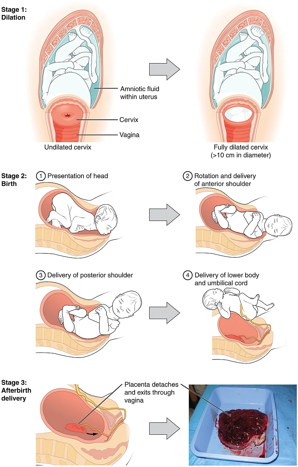
- First stage: from the onset of established labour to full dilatation; the latent phase precedes it and established labour is diagnosed from around 4 cm with regular painful contractions.
- Second stage: from full dilatation to birth, divided into a passive phase and an active pushing phase.
- Third stage: from birth of the baby to expulsion of the placenta and membranes; prolonged beyond 30 minutes with active management or 60 minutes without.
- Cardinal movements: engagement and descent, flexion, internal rotation, extension, restitution, external rotation and expulsion.
- Station: the level of the presenting part relative to the ischial spines, from -3 above to +3 below; station 0 means the head is engaged.
- Monitoring: intermittent auscultation every 15 minutes in the first stage and every 5 minutes in the second stage for low-risk women; continuous CTG if risk factors are present.
- Third stage management: active management with 10 IU oxytocin intramuscularly, deferred cord clamping and controlled cord traction reduces postpartum haemorrhage.
Introduction
Labour is the process by which regular, painful uterine contractions produce progressive effacement and dilatation of the cervix, leading to expulsion of the fetus, placenta and membranes. Normal labour is usually defined as spontaneous in onset, between 37+0 and 41+6 weeks, with a single fetus in a cephalic vertex presentation, progressing without intervention to a spontaneous vaginal birth.1
Understanding what is normal is the prerequisite for recognising what is not. Almost every obstetric emergency is defined by reference to normal labour: delay in the first stage, delay in the second stage, malposition, malpresentation, fetal compromise and postpartum haemorrhage are all deviations from the sequence described here.
Physiology of the onset of labour
No single trigger initiates labour. The prevailing view is that the myometrium is actively held quiescent throughout pregnancy, principally by progesterone, and that labour begins when a series of changes overcomes that inhibition.
- Fetal signal - maturation of the fetal hypothalamic-pituitary-adrenal axis raises fetal cortisol, which drives placental corticotrophin-releasing hormone in a positive feedback loop
- Functional progesterone withdrawal - a shift in the ratio of oestrogen to progesterone activity, mediated at receptor level rather than by falling circulating progesterone in humans
- Oxytocin receptor upregulation - myometrial oxytocin receptor density rises dramatically near term, so the uterus becomes responsive to circulating oxytocin rather than oxytocin levels rising
- Prostaglandins - PGE2 and PGF2-alpha ripen the cervix by degrading collagen and increasing water content, and stimulate contractions
- Gap junctions - connexin-43 gap junctions form between myocytes, allowing the uterus to contract as a coordinated syncytium rather than as isolated cells
- The Ferguson reflex - stretching of the cervix and upper vagina triggers a neuroendocrine reflex release of oxytocin, providing positive feedback
Uterine contractions in labour are unusual among smooth muscle contractions in showing retraction: after each contraction the muscle fibres of the upper segment do not return to their original length. This progressively shortens the upper segment, pulls the lower segment and cervix upwards over the presenting part, and is the mechanism of effacement and dilatation.
The first stage
The first stage runs from the onset of labour to full cervical dilatation at 10 cm. It is divided into a latent and an established phase, and the distinction matters because women admitted in the latent phase have higher rates of intervention.
| Phase | Definition | Typical duration |
|---|---|---|
| Latent | Painful contractions with cervical effacement and dilatation up to around 4 cm; contractions are often irregular | Highly variable, commonly 6-20 hours in a first labour; may be several days |
| Established (active) | Regular painful contractions with progressive dilatation from around 4 cm | Around 8 hours in a nulliparous woman and 5 hours in a parous woman, rarely exceeding 18 and 12 hours respectively |
NICE defines delay in the established first stage as dilatation of less than 2 cm in 4 hours in a nulliparous woman, or less than 2 cm in 4 hours or a slowing of progress in a parous woman. Other features suggesting delay are descent and rotation of the head failing to progress, and a change in the strength, duration or frequency of contractions.1
The mechanism of labour
The fetal head must negotiate a bony pelvis whose widest diameter changes orientation between the inlet and the outlet. At the pelvic inlet the transverse diameter is greater than the anteroposterior; at the outlet the reverse is true. The fetal head is itself oval, with a longer anteroposterior than transverse diameter, so it must rotate as it descends. This is the entire explanation for the cardinal movements.
| Movement | What happens and why |
|---|---|
| Engagement and descent | The widest transverse diameter of the head passes the pelvic inlet, usually in the occipito-transverse position; measured clinically as fifths palpable abdominally |
| Flexion | Pressure from the pelvic floor flexes the head onto the chest, presenting the smallest suboccipitobregmatic diameter of about 9.5 cm instead of the larger occipitofrontal diameter |
| Internal rotation | The occiput rotates anteriorly through 90 degrees as it meets the gutter of the levator ani, bringing the head into the occipito-anterior position to fit the anteroposterior outlet |
| Extension | The head extends as it passes under the pubic symphysis and the perineum distends; crowning occurs when the head no longer recedes between contractions |
| Restitution | The head untwists 45 degrees to realign with the shoulders, which remain oblique |
| External rotation | The shoulders rotate into the anteroposterior diameter of the outlet and the head turns a further 45 degrees to face laterally |
| Expulsion | The anterior shoulder delivers under the pubic symphysis, then the posterior shoulder over the perineum by lateral flexion, followed by the body |
The second stage
The second stage runs from full dilatation to the birth of the baby. It is divided into a passive phase, in which the cervix is fully dilated but there is no involuntary expulsive urge, and an active phase, in which the woman has an expulsive urge or is actively pushing.
| Parity | Birth expected within | Delay diagnosed after |
|---|---|---|
| Nulliparous | 3 hours of active second stage | 2 hours of active pushing |
| Parous | 2 hours of active second stage | 1 hour of active pushing |
Allowing an hour of passive second stage before active pushing in a nulliparous woman with an epidural permits the head to descend and rotate passively, and reduces the rate of instrumental delivery. Women should be encouraged to adopt whatever position they find comfortable and discouraged from lying supine, which reduces uteroplacental perfusion by aortocaval compression.
- Support the perineum and control the speed of the head at crowning to reduce trauma; warm compresses reduce third- and fourth-degree tears
- Episiotomy is not performed routinely; it is indicated for fetal compromise requiring expedited birth or for instrumental delivery, and should be mediolateral at 60 degrees from the midline
- Check for a nuchal cord after the head delivers, and either slip it over the head or, rarely, clamp and cut it
- Await restitution and external rotation, then apply gentle downward traction to deliver the anterior shoulder followed by upward traction for the posterior shoulder
- Note the time of birth and place the baby skin to skin with the mother
Monitoring in labour
Observations in a low-risk labour are deliberately minimal, with the intention of avoiding intervention that is not indicated.
| Observation | Frequency |
|---|---|
| Fetal heart by intermittent auscultation | Every 15 minutes in the first stage, for a full minute immediately after a contraction; every 5 minutes in the second stage |
| Maternal pulse | Hourly, and alongside every auscultation to distinguish maternal from fetal heart rate |
| Blood pressure and temperature | Every 4 hours |
| Contractions | Assess frequency every 30 minutes |
| Vaginal examination | Every 4 hours, or if there is concern about progress |
| Urine output and urinalysis | Encourage voiding and document; test for ketones and protein |
Continuous cardiotocography is recommended where there are antenatal or intrapartum risk factors: suspected chorioamnionitis or sepsis, severe hypertension, oxytocin augmentation, significant meconium, fresh vaginal bleeding, or an abnormality detected on intermittent auscultation. Offering continuous monitoring to low-risk women increases the caesarean and instrumental delivery rate without reducing cerebral palsy.
Reading a cardiotocograph
The standard structured approach is summarised as DR C BRAVADO: Define Risk, Contractions, Baseline RAte, Variability, Accelerations, Decelerations, Overall impression. A normal baseline is 110-160 beats per minute with variability of 5-25 beats per minute. Early decelerations mirror contractions and reflect head compression. Variable decelerations vary in timing and shape and reflect cord compression. Late decelerations begin after the peak of the contraction and recover after it ends, and indicate uteroplacental insufficiency. Features are classified as reassuring, non-reassuring or abnormal, and the trace overall as normal, suspicious or pathological.2
Analgesia in labour
| Method | Notes |
|---|---|
| Continuous support, mobility, position change | One-to-one midwifery support reduces the need for analgesia and improves satisfaction |
| Immersion in water | Effective for pain in the first stage; contraindicated where continuous monitoring is required |
| TENS | May help in early labour; little evidence of benefit in established labour |
| Entonox (50% nitrous oxide in oxygen) | Rapid onset and offset, self-administered; causes light-headedness and nausea but no fetal depression |
| Intramuscular opioids (pethidine, diamorphine) | Modest analgesia with significant sedation and nausea; crosses the placenta, causing neonatal respiratory depression and drowsiness that may delay feeding; give an antiemetic |
| Remifentanil patient-controlled analgesia | Short-acting; requires one-to-one care and continuous oxygen saturation monitoring because of apnoea risk |
| Epidural | The most effective analgesia available; a low-dose local anaesthetic and opioid combination is used |
Women should be counselled accurately about epidurals. They provide much better pain relief than any alternative, and there is no evidence that they increase the caesarean section rate or cause long-term backache. They do prolong the second stage and increase the rate of instrumental vaginal birth, require intravenous access and more intensive monitoring, and cause hypotension, urinary retention and pruritus. Accidental dural puncture occurs in around 1 in 100 insertions and causes a postural headache in the majority of those affected.1
The third stage
The third stage runs from birth of the baby to expulsion of the placenta and membranes. Signs of placental separation are a gush of blood, lengthening of the cord and the uterus rising and becoming globular. It is prolonged if not completed within 30 minutes with active management, or within 60 minutes with physiological management.
| Element | Active management | Physiological management |
|---|---|---|
| Uterotonic | Oxytocin 10 IU intramuscularly with birth of the anterior shoulder or immediately after birth; Syntometrine may be used if there is no hypertension | None given |
| Cord clamping | Deferred for at least 1 minute, and up to 5 minutes, unless the baby needs resuscitation | Cord clamped after pulsation ceases |
| Delivery of placenta | Controlled cord traction with counter-traction on the uterus, once the uterus is contracted | Maternal effort alone |
| Postpartum haemorrhage risk | Around 5% | Around 15% |
Active management is recommended for all women, but physiological management is a reasonable choice for a woman at low risk who requests it, with a clear plan to convert to active management if there is bleeding, if the placenta is not delivered within an hour, or if the woman wishes. Never apply controlled cord traction without a uterotonic and without a contracted uterus, because of the risk of uterine inversion.1,3
Immediately after birth
- Skin-to-skin contact immediately and uninterrupted where possible; it improves thermoregulation, breastfeeding initiation and bonding
- Apgar score at 1 and 5 minutes, assessing appearance, pulse, grimace, activity and respiration
- Vitamin K offered to all babies, intramuscularly as a single dose or orally in a course, to prevent haemorrhagic disease of the newborn
- First feed within the first hour where possible, with support for breastfeeding
- Examine the placenta and membranes for completeness; retained fragments cause haemorrhage and infection
- Estimate blood loss by weighing swabs and measuring, rather than by visual estimation, which systematically underestimates
- Palpate the uterine fundus to confirm it is contracted, and inspect the perineum, vagina and cervix for trauma
- Newborn and infant physical examination within 72 hours, and the newborn blood spot screen at day 5
Perineal trauma
| Degree | Structures involved | Management |
|---|---|---|
| First | Vaginal epithelium or perineal skin only | May not need suturing if haemostatic and well apposed |
| Second | Perineal muscles, anal sphincter intact | Suture; includes an episiotomy |
| Third (3a, 3b, 3c) | Anal sphincter complex: 3a is under 50% of the external sphincter, 3b over 50%, 3c involves the internal sphincter | Repair in theatre by a trained clinician, with antibiotics, laxatives and physiotherapy follow-up |
| Fourth | Anal sphincter complex and anorectal mucosa | Repair in theatre; long-term follow-up for incontinence |
When labour is not normal
Failure to progress is traditionally analysed in terms of the three Ps. The powers may be inadequate, with hypotonic or poorly coordinated contractions, which is the commonest cause in a nulliparous woman and is treated with amniotomy and oxytocin augmentation. The passenger may be malpositioned, most often occipito-posterior, malpresenting, or simply large. The passages may be inadequate, either because of pelvic shape or, rarely, because of a soft tissue obstruction such as a cervical fibroid.
In a parous woman, poor progress should never be attributed to inadequate powers without careful thought, because a uterus that has already delivered a baby is usually capable of doing so again. Delay in a multipara raises the possibility of obstruction, malposition or malpresentation, and carries a real risk of uterine rupture if oxytocin is used uncritically.1
The partogram is the tool that makes deviation visible. Plotting cervical dilatation against time, alongside descent, contractions, fetal heart rate, liquor and maternal observations, converts a series of individual assessments into a trend, and is what prompts the question of whether labour is progressing normally at the point at which something can still be done about it.
References
- NICE CG190. Intrapartum care for healthy women and babies. 2014 (updated 2023). Available here
- NICE NG229. Fetal monitoring in labour. 2022. Available here
- RCOG Green-top Guideline No. 52. Prevention and management of postpartum haemorrhage. Available here
This article is written for revision and education. It is not clinical guidance and must not be used to make decisions about the care of a patient. Always check current NICE guidance and local protocols.