
Fractures: Principles of Assessment and Management
Key points
- Fracture: a break in the structural continuity of bone, ranging from a hairline crack to complete separation with displacement.
- Open vs closed: an open fracture communicates with a breach in the overlying skin - it is a surgical emergency because of the infection risk, not because it is necessarily more displaced.
- Neurovascular status: document distal pulses, capillary refill, sensation and motor function before and after every manipulation - this is the single most medicolegally important part of the examination.
- Open fracture emergency care: analgesia, a single photograph then cover the wound, realign and splint, IV antibiotics within 1 hour, check tetanus status, urgent combined orthoplastic referral.
- Imaging: plain radiographs in two orthogonal views, including the joint above and below the injury; CT for complex or intra-articular fractures; MRI for occult fractures such as scaphoid or femoral neck.
- Management principle: reduce, hold, rehabilitate - closed or open reduction, then a cast, ORIF, intramedullary nail or external fixator to hold it, then physiotherapy.
- Healing: most fractures heal by secondary (callus) healing through haematoma, soft callus, hard callus and remodelling; rigid fixation with no gap allows primary healing without callus.
- Pathological fracture: suspect this when the fracture is out of proportion to the trauma, or preceded by unexplained bone pain - osteoporosis is the commonest cause, metastasis the one not to miss.
Introduction
A fracture is a break in the structural continuity of bone. This includes complete fractures, incomplete fractures where the bone is cracked or bent but not fully separated, and microscopic fatigue damage from repetitive loading that never involves a single traumatic event at all. The term covers everything from an undisplaced hairline crack in a metatarsal to a shattered femoral shaft after a road traffic collision.
Fractures are extremely common in UK clinical practice, seen in every emergency department, every trauma call and a large proportion of orthopaedic outpatient referrals. This article covers the general principles that apply to fractures anywhere in the skeleton: how they are classified, how they present, how to examine a limb safely, how open fractures are managed as an emergency, how they are imaged, the reduce-hold-rehabilitate framework for treatment, and how bone actually heals.
It deliberately does not go deep into any single fracture site - the neck of femur, for example, has its own dedicated article, as do the complications that can follow any fracture and compartment syndrome specifically. Treat this as the scaffolding that those more specific topics hang off.
Classification
Fractures are classified along several independent axes, and exam questions frequently test more than one at once - for example, an open, comminuted, pathological fracture of the femur ticks three separate boxes.
Open versus closed
A closed fracture has intact overlying skin. An open (compound) fracture communicates with a wound through the skin, whether from the bone piercing outwards or from the same force that broke the bone also breaching the skin from outside. Open fractures are managed as surgical emergencies primarily because of the risk of deep infection and osteomyelitis, not because they are necessarily more severe or displaced than a closed injury - a small puncture wound over a minimally displaced tibial fracture is still an open fracture.
Complete versus incomplete
- Complete fracture - the bone is broken into two or more separate fragments across its full width
- Incomplete fracture - the bone is cracked or deformed but remains in continuity, seen almost exclusively in children because their bone is more porous and the periosteum thicker and stronger
- Greenstick fracture - one cortex breaks while the opposite cortex bends, like snapping a green twig; typically diaphyseal long bone fractures in young children
- Buckle (torus) fracture - a compression failure of the cortex, which bulges outwards without a visible fracture line; classically the distal radius in a child who has fallen on an outstretched hand, and often manageable in a removable splint alone
Fracture pattern
The pattern of the fracture line reflects the mechanism of injury, which is worth reading off the radiograph before you even look at the history.
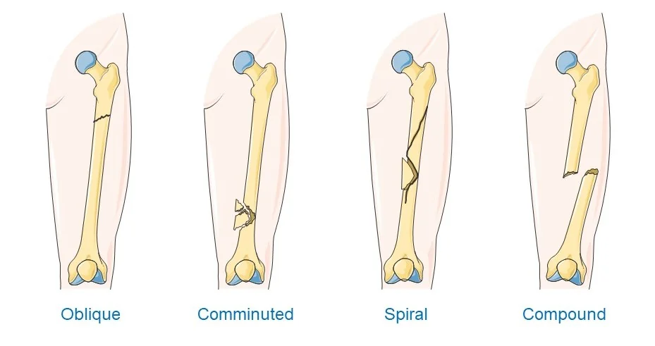
| Pattern | Typical mechanism | Notes |
|---|---|---|
| Transverse | Direct blow, or a pure bending force | Fracture line runs at right angles to the long axis of the bone |
| Oblique | Bending force with an axial component | Fracture line runs at an angle across the bone |
| Spiral | Torsional (rotational) force | Long, curving fracture line; a spiral fracture in a child who is not yet mobile should prompt consideration of non-accidental injury |
| Comminuted | High-energy trauma | More than two fragments; associated with more soft tissue damage and a higher risk of non-union |
| Segmental | High-energy trauma | Two separate fracture lines isolate a segment of shaft, which can have a precarious blood supply |
| Avulsion | Sudden traction through a tendon or ligament | A fragment of bone is pulled off at the attachment site, e.g. the tibial tuberosity or the base of the fifth metatarsal |
Stress fractures
A stress (fatigue) fracture occurs in structurally normal bone that has been subjected to repetitive, submaximal loading without adequate time to remodel, rather than a single traumatic event. Classic examples are the second metatarsal in military recruits after long marches ("march fracture") and the tibia or navicular in runners. They present with gradually worsening, activity-related pain that is often absent or subtle on the initial radiograph, and MRI or isotope bone scan may be needed to confirm the diagnosis.
Pathological fractures
A pathological fracture occurs through bone that is already weakened by an underlying disease process, so that a force which would not break normal bone is sufficient to cause a fracture. Osteoporosis is by far the commonest cause and underlies the majority of fragility fractures in older adults, particularly of the distal radius, proximal femur and vertebral bodies. Other causes to consider are bone metastases (breast, lung, prostate, kidney and thyroid account for most), primary bone tumours, myeloma, osteomyelitis, Paget's disease, and metabolic bone disease such as osteomalacia.
Mechanism of injury
Understanding how a fracture occurred explains its pattern, predicts associated soft tissue and organ injury, and guides how urgently it needs to be treated.
- Direct trauma - a force applied straight to the bone, such as a blow from a blunt object or a bumper injury to the tibia, typically producing a transverse or comminuted fracture at the point of impact
- Indirect trauma - force transmitted along the bone or through a joint from a distant point of impact, for example a fall on an outstretched hand transmitting force up the forearm to fracture the clavicle or proximal humerus
- High-energy mechanisms - road traffic collisions, falls from height, industrial and sporting trauma; associated with comminution, extensive soft tissue damage, open wounds and a higher likelihood of life-threatening associated injuries, so these patients need a full trauma assessment, not just a look at the injured limb
- Low-energy mechanisms - a fall from standing height, particularly in an older person; the fracture itself may look unremarkable, but a low-energy fracture in this group should always trigger a falls assessment and consideration of underlying osteoporosis
- Repetitive submaximal loading - the mechanism behind stress fractures, discussed above
- Pathological weakening - osteoporosis, malignancy or infection reducing the force required to fracture the bone, discussed above
Clinical features
The classic features of a fracture are pain, swelling, deformity, bruising and inability to use or weight-bear through the affected limb, but not all are present in every case - an undisplaced or incomplete fracture may show only localised tenderness and reluctance to move the limb.
- Pain - typically severe, worse on any attempted movement, and localised to the fracture site, though referred pain can mislead (hip pathology presenting as knee pain in children, for example)
- Swelling - from haematoma and soft tissue oedema, often delayed by minutes to hours after the injury
- Deformity - visible angulation, shortening or rotation of the limb
- Bruising - may take hours to become visible, and can track along fascial planes away from the fracture site
- Inability to weight-bear or use the limb - a child who refuses to weight-bear after a fall has a fracture until proven otherwise
- Crepitus - a grating sensation or sound from fracture fragments moving against each other; do not deliberately elicit this, as it is painful and can worsen soft tissue or neurovascular injury
- Point tenderness - the single most sensitive sign on examination, and often the only positive finding in an undisplaced fracture
Examination
Examine every suspected fracture with the standard look, feel, move structure, always comparing with the uninjured side, and always assessing the joint above and below the injury as well as the injury itself.
- Look - deformity, swelling, bruising, skin colour and integrity, any open wounds or skin tenting from an underlying fragment, and the position the limb is being held in
- Feel - point tenderness, warmth, and, critically, the distal neurovascular examination described below
- Move - active movement first if the patient is able, then gentle passive movement; do not force movement through resistance, and stop immediately if this reproduces severe pain
Neurovascular assessment
Distal neurovascular status must be assessed and clearly documented before and after any manipulation or reduction, and again after any splint or cast is applied, because a splint that is too tight can itself cause vascular or nerve compromise. This is tested repeatedly in OSCEs and is one of the most common sources of missed pathology and litigation in real practice.
- Pulses - palpate the relevant distal pulse (e.g. radial, dorsalis pedis and posterior tibial) and compare with the other side
- Capillary refill time - should be under 2 seconds in the digits distal to the injury
- Colour and temperature - a pale, cold limb suggests critical ischaemia
- Sensation - test light touch in the distribution of each nerve at risk for that particular fracture, not just "sensation intact"
- Motor function - test the specific muscle groups supplied by each nerve at risk, again comparing with the other side
Open fractures
Open fractures are surgical emergencies because the wound gives bacteria direct access to bone, with a real risk of deep infection, chronic osteomyelitis and non-union if management is delayed or substandard. The Gustilo-Anderson classification, though originally intra-operative and now often criticised for its poor inter-observer reliability, is still widely used to describe severity and to communicate between teams.1
| Type | Wound | Soft tissue damage | Contamination / energy |
|---|---|---|---|
| I | Less than 1 cm | Minimal | Clean, low energy, simple fracture pattern |
| II | 1-10 cm | Moderate | Moderate energy, moderate contamination |
| IIIA | Usually over 10 cm | Extensive, but adequate soft tissue for coverage of bone | High energy; comminuted or segmental fractures |
| IIIB | Usually over 10 cm | Extensive soft tissue loss with periosteal stripping | Bone exposed, requires flap coverage; heavily contaminated |
| IIIC | Any size | As above | Associated arterial injury requiring repair - always type IIIC regardless of wound size |
Emergency management
UK practice follows BOAST (British Orthopaedic Association Standards for Trauma) guidance on the initial management of open fractures.2 After the usual ATLS-style primary survey and resuscitation of any polytrauma patient, the sequence for the injured limb is:
- Analgesia - given early and adequately, following the WHO pain ladder or with IV opioids for severe pain; do not let ongoing pain assessment be delayed by imaging
- Photograph the wound once, then cover it with a saline-soaked dressing and leave it alone - repeated exposure in the emergency department for looking or re-photographing only increases infection risk
- Realign and splint the limb, restoring length and correcting gross deformity; this reduces ongoing soft tissue and neurovascular injury and improves pain
- Reassess and document distal neurovascular status before and after realignment
- IV antibiotics as soon as possible, ideally within an hour of injury, per local protocol - typically co-amoxiclav or cefuroxime, with the addition of gentamicin for heavily contaminated wounds (e.g. agricultural or aquatic injuries), and teicoplanin or vancomycin substituted for penicillin allergy3
- Check tetanus immunisation status and give tetanus-containing vaccine and/or tetanus immunoglobulin according to the wound category and vaccination history4
- Urgent referral for combined orthoplastic care - definitive debridement is planned jointly by orthopaedic and plastic surgery teams, usually within 12-24 hours of injury, sooner for heavily contaminated or marine/agricultural wounds
Investigations
Plain radiographs are the first-line investigation for almost all suspected fractures, and should always include two orthogonal views - typically anteroposterior and lateral - since a fracture or the degree of displacement can be entirely hidden on a single view. The film should also include the joint above and below the injury, both to identify a second, less obvious fracture or dislocation and because management often depends on involvement of the adjacent joint.
- CT - used for complex or intra-articular fractures where the three-dimensional anatomy affects surgical planning, for example tibial plateau, calcaneal, pelvic and complex peri-articular fractures, and for fractures in anatomically complex regions such as the spine and pelvis where plain films are hard to interpret
- MRI - the investigation of choice for a suspected fracture that is clinically obvious but not visible on plain radiographs, most classically a scaphoid fracture with a normal initial X-ray, or a suspected femoral neck fracture in an older patient with hip pain and a normal-looking film. Also used to characterise stress fractures and to assess soft tissue and ligamentous injury alongside bone
- Isotope bone scan - an alternative to MRI for occult or stress fractures where MRI is unavailable or contraindicated, though it is less specific and slower to arrange
- Ultrasound - a useful bedside adjunct in children for suspected forearm buckle fractures and in some rib fracture assessments, reducing radiation exposure
Principles of management
Once any immediately life- or limb-threatening issue has been addressed, fracture management follows the same broad framework regardless of site: reduce, hold, rehabilitate.
Analgesia
Adequate analgesia should be given from the first assessment, following the WHO analgesic ladder and escalating as needed - paracetamol and NSAIDs for mild pain, weak opioids for moderate pain, and titrated IV opioids for severe pain or before any manipulation. Reduction of a displaced fracture is itself painful and is usually performed under procedural sedation, regional nerve or haematoma block, or general anaesthesia, depending on the fracture, the patient, and local facilities.
Reduce
Reduction means restoring the fracture fragments to (or close to) their normal anatomical alignment.
- Closed reduction - manipulating the limb externally, under sedation, regional block or general anaesthesia, without surgically exposing the fracture; suitable for many extra-articular and minimally displaced fractures
- Open reduction - surgically exposing the fracture site to reduce it under direct vision, needed when closed reduction fails, when the fracture is intra-articular and requires anatomical restoration of the joint surface, or when there are multiple fragments that cannot be aligned closed
Hold
Once reduced (or if the fracture is already acceptably aligned), it must be held in position while it heals. The choice of method depends on the fracture pattern, its stability, the bone involved, the patient's physiology and functional demands, and whether the fracture is open.
| Method | Typical use | Notes |
|---|---|---|
| Cast or splint immobilisation | Stable, minimally displaced or successfully closed-reduced fractures | Non-invasive; requires monitoring for tight cast/compartment syndrome and joint stiffness with prolonged immobilisation |
| Open reduction and internal fixation (ORIF) with plates and screws | Intra-articular fractures needing anatomical reduction; fractures near joints; fractures that fail closed reduction | Allows precise reduction under direct vision; carries surgical risks of infection, wound healing problems and metalwork failure |
| Intramedullary nailing | Diaphyseal long bone fractures, e.g. femoral and tibial shaft | Load-sharing device inserted through the medullary canal; generally allows earlier weight-bearing than a plate |
| External fixation | Damage control in open, heavily contaminated or unstable fractures, and in some pelvic fractures | Pins through the skin into bone connected by an external frame; allows access to soft tissue for wound care and can be temporary before conversion to internal fixation, or definitive in selected cases |
Rehabilitate
Rehabilitation begins as soon as the fracture is adequately held and is not an afterthought - it is what determines whether the patient returns to their previous level of function. This includes physiotherapy to restore range of movement and strength, occupational therapy input for activities of daily living, clear weight-bearing instructions specific to the fixation used, and, in frail or older patients, comprehensive geriatric assessment and orthogeriatric input to address falls risk, bone health and comorbidities in parallel with the fracture itself.
Fracture healing
Most fractures heal by secondary (indirect) bone healing, which proceeds through a well-defined biological sequence, though the exact timescale varies enormously with the bone involved, the patient's age and how well the fracture is immobilised.
- Haematoma formation (hours to days) - bleeding from the fracture ends and surrounding soft tissue forms a clot, and the resulting inflammatory response, with cytokines and growth factors, recruits the cells needed for repair
- Soft (fibrocartilaginous) callus (roughly days to a few weeks) - fibroblasts and chondroblasts lay down a soft matrix of collagen and cartilage bridging the fracture gap, providing early, if flimsy, stability
- Hard (bony) callus (roughly weeks to a few months) - the soft callus is converted to woven bone through endochondral ossification, producing a bulky but mechanically sound union across the fracture
- Remodelling (months to years) - the disorganised woven bone is gradually replaced by organised lamellar bone and reshaped along the lines of mechanical stress, following Wolff's law, eventually restoring something close to the bone's original shape and strength
Primary (direct) bone healing is different and only occurs when a fracture is rigidly fixed with anatomical reduction and essentially no gap between the fragments, as with compression plating. Here, osteons cross the fracture line directly via so-called cutting cones, without ever forming a callus. It is a slower, more fragile process biologically and depends entirely on rigid, stable fixation, which is why callus on a radiograph after plate fixation can actually be a sign that the construct is not rigid enough.
Factors that impair healing
- Smoking - nicotine impairs blood flow to the fracture site and is one of the strongest modifiable risk factors for delayed and non-union
- NSAIDs - inhibit prostaglandin-mediated bone formation, and are generally avoided in the early weeks after a fracture, particularly where union is already a concern
- Diabetes mellitus - impairs microvascular supply and is associated with delayed union and higher infection rates
- Poor local blood supply - certain sites are notoriously prone to non-union or avascular necrosis because of their precarious vascular anatomy, including the scaphoid waist and proximal pole, the femoral neck, and the talus
- Infection - directly disrupts the healing cascade and is a particular risk after open fractures or surgical fixation
- Inadequate immobilisation - excessive movement at the fracture site disrupts the soft and hard callus as it forms
- Patient age - children heal considerably faster than adults, and healing slows further with advancing age
- Nutritional status and corticosteroid use - malnutrition, vitamin D deficiency and long-term steroids all impair bone formation and remodelling
Complications
This is only a brief overview - the full range of fracture complications, and how to recognise and manage each of them, is covered in the dedicated Complications of Fractures article, and compartment syndrome specifically has its own article given how frequently it is examined and how easily it is missed.
- Early/immediate: neurovascular injury, compartment syndrome, fat embolism syndrome (classically after long bone or pelvic fractures), haemorrhage and hypovolaemic shock, associated visceral injury
- Late: delayed union and non-union, malunion, avascular necrosis, infection including chronic osteomyelitis, joint stiffness and contracture, complex regional pain syndrome, post-traumatic osteoarthritis
Red flags
References
- Gustilo RB, Anderson JT. Prevention of infection in the treatment of one thousand and twenty-five open fractures of long bones. Journal of Bone and Joint Surgery (Am). 1976. Available here
- British Orthopaedic Association. BOAST - Open Fractures. Standards for Trauma. Available here
- BNF. Antibacterial prophylaxis - surgical and trauma wounds. Available here
- UK Health Security Agency. Tetanus: the green book, chapter 30. Available here
- NICE NG37. Fractures (non-complex): assessment and management. 2016. Available here
- NICE NG38. Fractures (complex): assessment and management. 2016. Available here
- NICE NG12. Suspected cancer: recognition and referral. 2015, updated 2023. Available here
- NICE CG124. Hip fracture: management. 2011, updated 2023. Available here
- Solomon L, Warwick D, Nayagam S. Apley and Solomon's System of Orthopaedics and Trauma. 10th ed. Available here
- Marsh JL, Slongo TF, Agel J et al. Fracture and dislocation classification compendium. Journal of Orthopaedic Trauma. 2007. Available here
This article is written for revision and education. It is not clinical guidance and must not be used to make decisions about the care of a patient. Always check current NICE guidance and local protocols.