
Myelodysplastic Syndromes
Key points
- Myelodysplastic syndromes (MDS): a group of clonal haematopoietic stem cell disorders causing dysplastic (abnormally formed), ineffective haematopoiesis, resulting in cytopenias despite a normal or hypercellular bone marrow - production is high but mostly non-productive.
- Epidemiology: predominantly a disease of the elderly (median age at diagnosis around 70). May be primary (de novo) or secondary/therapy-related, following previous chemotherapy or radiotherapy - the latter carries a worse prognosis.
- The central risk: MDS is a pre-leukaemic condition: a variable proportion of patients progress to acute myeloid leukaemia, and risk of transformation is central to prognosis and treatment decisions.
- Clinical features: insidious symptoms of one or more cytopenias - fatigue (anaemia), infection (neutropenia), bruising/bleeding (thrombocytopenia) - often in an older patient with no other obvious cause.
- Diagnosis: FBC cytopenias with dysplastic blood film features, confirmed by bone marrow aspirate and trephine showing dysplasia across one or more lineages, plus cytogenetics, which is essential for both diagnosis and prognosis.
- Risk stratification: the Revised International Prognostic Scoring System (IPSS-R) combines cytopenia severity, blast percentage and cytogenetics to stratify patients from very low to very high risk, and drives the treatment approach.
- Management: ranges from supportive care and growth factors in low-risk disease, through azacitidine for higher-risk disease, to allogeneic stem cell transplant - the only curative option - in selected fit, higher-risk patients.
- 5q- syndrome: a distinct, favourable-prognosis MDS subtype with an isolated deletion of chromosome 5q, characteristically responsive to lenalidomide.
Introduction
Myelodysplastic syndromes are clonal disorders of the haematopoietic stem cell in which blood cell precursors mature abnormally (dysplasia) and are destroyed within the marrow before release, or are released as poorly functioning cells - a state of ineffective haematopoiesis. The result is one or more peripheral blood cytopenias despite a bone marrow that is often normo- or hypercellular.1
MDS matters clinically for two reasons: it causes significant morbidity through the cytopenias themselves, and it is a pre-leukaemic state, with a variable but ever-present risk of transformation to acute myeloid leukaemia (AML).
Aetiology
- Primary (de novo) MDS - the majority of cases, with no identifiable prior cause, strongly associated with increasing age
- Secondary (therapy-related) MDS - following previous cytotoxic chemotherapy (particularly alkylating agents and topoisomerase II inhibitors) or radiotherapy, typically presenting years after treatment and carrying a worse prognosis and higher-risk cytogenetics than de novo disease
- Rare predisposing conditions include prior exposure to benzene and other marrow toxins, and certain inherited bone marrow failure syndromes
Clinical features
Onset is typically insidious, and many cases are picked up incidentally on a routine blood count in an older patient. Symptoms reflect whichever cell line is most affected:
- Anaemia - the commonest presentation; fatigue, dyspnoea, pallor
- Neutropenia - recurrent or unusual infections
- Thrombocytopenia - easy bruising, petechiae, mucosal bleeding
- Some patients have more than one cytopenia at presentation; a minority have only an isolated abnormality (e.g. macrocytosis) noted incidentally
- Hepatosplenomegaly and lymphadenopathy are not typical of MDS and should prompt consideration of an alternative or overlapping diagnosis
Investigations
Full blood count and blood film
Cytopenias affecting one or more lineages, often with a macrocytosis even in the absence of B12/folate deficiency. The blood film may show dysplastic features:
- Hypogranular or hyposegmented neutrophils ("pseudo-Pelger-Huet" cells, with a bilobed rather than multi-lobed nucleus)
- Giant or agranular platelets
- Basophilic stippling, and circulating blasts in higher-risk disease
Bone marrow examination - the key diagnostic test
Bone marrow aspirate and trephine biopsy is required for diagnosis, showing dysplastic changes across one or more of the erythroid, myeloid and megakaryocytic lineages, and allows the blast percentage to be counted (which distinguishes MDS, where blasts are <20%, from acute leukaemia, where they are ≥20%).1
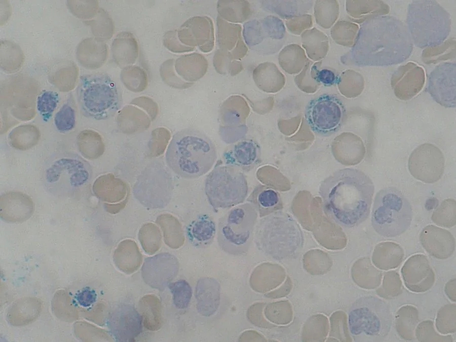
Perls' (Prussian blue) staining identifies ring sideroblasts - erythroid precursors with iron-laden mitochondria forming a ring around the nucleus - which define specific MDS subtypes (MDS with ring sideroblasts) and reflect defective haem synthesis.
Cytogenetics
Essential for both diagnosis and prognosis. Common abnormalities include deletions of chromosome 5q, 7q and 20q, monosomy 7, and complex karyotypes (which carry a particularly poor prognosis). The isolated 5q- deletion defines a distinct, favourable-risk subtype.
Excluding mimics
B12 and folate levels, copper level, and HIV testing should be checked, since severe deficiency states and HIV can cause dysplastic changes that mimic MDS but are reversible with treatment of the underlying cause.
Risk stratification and classification
The Revised International Prognostic Scoring System (IPSS-R) combines the degree of cytopenia, bone marrow blast percentage, and cytogenetic risk group to stratify patients into very low, low, intermediate, high and very high risk categories, which directly guides management intensity and estimates median survival and risk of progression to AML.2
The WHO classification subdivides MDS by the number of dysplastic lineages, blast percentage, and specific genetic features (e.g. MDS with ring sideroblasts, MDS with isolated del(5q)), refining prognosis further.
Differential diagnosis
- Severe B12 or folate deficiency - causes megaloblastic, dysplastic-appearing changes that resolve with replacement; always exclude before diagnosing MDS
- Aplastic anaemia - hypocellular marrow without dysplasia, rather than the normo/hypercellular dysplastic marrow of MDS
- Acute myeloid leukaemia - distinguished by a bone marrow blast count of 20% or more
- Other causes of cytopenia - hypersplenism, marrow infiltration, drug effect, HIV
- Myeloproliferative/myelodysplastic overlap syndromes (e.g. chronic myelomonocytic leukaemia), which combine proliferative and dysplastic features
Management
Lower-risk disease
- Supportive care - blood and platelet transfusion as needed, with iron chelation if transfusion becomes frequent
- Erythropoiesis-stimulating agents for anaemia in selected patients with a low endogenous EPO level
- Lenalidomide - particularly effective in MDS with isolated del(5q), often achieving transfusion independence
- Growth factors (G-CSF) used judiciously for severe, symptomatic neutropenia
Higher-risk disease
- Hypomethylating agents (azacitidine) - standard first-line therapy for higher-risk MDS not proceeding straight to transplant, improving survival and delaying progression to AML3
- Allogeneic haematopoietic stem cell transplant - the only curative option, reserved for fit patients (fitness matters more than chronological age alone) with higher-risk disease and a suitable donor
- Clinical trial enrolment where available, given the rapidly evolving treatment landscape
Complications
- Transformation to acute myeloid leukaemia - the central long-term risk, with likelihood determined by IPSS-R risk group
- Infection, from neutropenia and also from qualitative neutrophil dysfunction even when the count is preserved
- Bleeding, from thrombocytopenia and qualitative platelet dysfunction
- Transfusion-related iron overload in transfusion-dependent patients
- Reduced quality of life from chronic fatigue and repeated hospital visits for monitoring and transfusion
Red flags
Prognosis
Prognosis in MDS is highly variable and driven predominantly by IPSS-R risk category: very low-risk disease can have a median survival exceeding 8 years with a low risk of AML transformation, while very high-risk disease carries a median survival of under a year without transplant and a high likelihood of progression to AML.2
Allogeneic stem cell transplant remains the only treatment that offers cure, but is limited by patient fitness, donor availability and the risks of the procedure itself, so most patients are managed with a combination of supportive care and disease-modifying therapy aimed at controlling cytopenias and delaying progression rather than cure.
References
- Cazzola M. Myelodysplastic Syndromes. N Engl J Med. 2020. Available here
- Greenberg PL, Tuechler H, Schanz J et al. Revised International Prognostic Scoring System for myelodysplastic syndromes. Blood. 2012. Available here
- Fenaux P, Mufti GJ, Hellstrom-Lindberg E et al. Efficacy of azacitidine compared with that of conventional care regimens in higher-risk MDS. Lancet Oncol. 2009. Available here
- British Society for Haematology. Guidelines for the diagnosis and management of adult myelodysplastic syndromes. Available here
- Paulo Henrique Orlandi Mourao, CC BY-SA 3.0, via Wikimedia Commons. Available here
This article is written for revision and education. It is not clinical guidance and must not be used to make decisions about the care of a patient. Always check current NICE guidance and local protocols.