
Quinsy (Peritonsillar Abscess)
Key points
- Quinsy: a peritonsillar abscess - a collection of pus between the tonsillar capsule and the superior constrictor muscle, usually complicating acute tonsillitis.
- Key features: severe unilateral throat pain, trismus, and a muffled "hot potato" voice, distinguishing it from simple bilateral tonsillitis.
- Examination: unilateral peritonsillar swelling with medial displacement of the tonsil and uvular deviation away from the affected side.
- Aetiology: polymicrobial, including Group A Streptococcus, Staphylococcus aureus and anaerobes such as Fusobacterium.
- Management: urgent ENT referral for needle aspiration or incision and drainage, plus IV antibiotics covering anaerobes.
- Adjuncts: corticosteroids are increasingly used alongside drainage and antibiotics to reduce pain and swelling.
- Complications: airway compromise, spread to the parapharyngeal or retropharyngeal space, and rarely Lemierre's syndrome or mediastinitis.
- Prognosis: excellent with prompt drainage; recurrence occurs in a minority, who may be offered interval tonsillectomy.
Introduction
Quinsy, or peritonsillar abscess, is a collection of pus in the potential space between the tonsillar capsule and the superior constrictor muscle. It is the most common deep neck space infection encountered in ENT practice and typically develops as a complication of acute tonsillitis that has progressed from cellulitis to a discrete abscess.1
It matters clinically because it looks similar to severe tonsillitis at first glance but requires a fundamentally different treatment - surgical drainage - and because, left untreated, infection can spread into the deep neck spaces and threaten the airway.
Aetiology and pathophysiology
Quinsy usually arises as a complication of acute tonsillitis, with infection spreading from the tonsil itself into the surrounding peritonsillar space. Infection is typically polymicrobial, involving aerobic organisms such as Group A Streptococcus and Staphylococcus aureus alongside anaerobes such as Fusobacterium necrophorum and Bacteroides species.1,2
Weber's glands - minor salivary glands located just above the tonsil - are thought to play a role in some cases, as their obstruction and infection can seed the adjacent peritonsillar space independently of the tonsil itself.
Risk factors
- Current or recent acute tonsillitis
- Smoking
- Poor dental hygiene and periodontal disease
- Young adults, typically 20s-40s, though it can occur at any age including in children
- Previous episode of quinsy
- Chronic tonsillitis
Clinical features
Patients typically report a few days of worsening sore throat that becomes markedly worse and unilateral, often out of proportion to what would be expected from tonsillitis alone. Key features include trismus (difficulty opening the mouth due to spasm of the pterygoid muscles), a muffled, "hot potato" voice, odynophagia severe enough to cause drooling, and referred pain to the ipsilateral ear.1
Systemic features - fever, malaise and halitosis - are common, and patients often look unwell. Neck stiffness or pain on moving the neck can occur if there is associated cervical lymphadenitis or early spread towards the parapharyngeal space.
Examination
Examination (as far as trismus allows) shows unilateral peritonsillar swelling, with the affected tonsil pushed medially and inferiorly, and the uvula deviated to the contralateral side. The soft palate on the affected side is typically swollen and erythematous.1
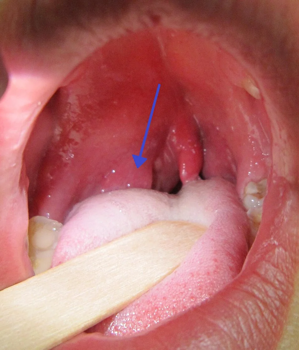
Cervical lymphadenopathy on the affected side is common and tender. Assess voice quality, ability to swallow secretions, and for any stridor, which would suggest impending airway compromise and needs immediate escalation rather than routine assessment.
Differential diagnosis
- Severe tonsillitis: bilateral swelling, no trismus and no uvular deviation
- Parapharyngeal abscess: deeper infection, often with more diffuse neck swelling and torticollis; may coexist with or follow untreated quinsy
- Retropharyngeal abscess: more common in young children, presenting with neck stiffness and reluctance to extend the neck
- Epiglottitis: drooling and stridor with a preference for sitting forward, but typically without the unilateral peritonsillar swelling of quinsy
- Dental or periodontal abscess: localised to the dentition rather than the peritonsillar space
Investigations
Quinsy is primarily a clinical diagnosis. FBC, CRP and blood cultures are useful in assessing severity, particularly if the patient looks systemically unwell.1 CT of the neck with contrast is reserved for cases with diagnostic uncertainty, or where deeper spread (parapharyngeal or retropharyngeal extension) is suspected, since it clearly delineates the extent of any collection. Pus obtained at drainage should be sent for microscopy, culture and sensitivity to guide any change in antibiotic therapy.
Management
Suspected quinsy warrants urgent same-day ENT referral. Definitive treatment is drainage of the abscess, most commonly by needle aspiration under local anaesthetic, which is both diagnostic (confirming pus) and therapeutic; incision and drainage is used if aspiration is unsuccessful or the collection is large.1,2
Intravenous antibiotics covering the polymicrobial flora, including anaerobes, are started alongside drainage - for example co-amoxiclav, or benzylpenicillin combined with metronidazole - with a switch to oral therapy once the patient is improving and able to swallow. IV fluids and analgesia are important given the odynophagia often limits oral intake, and a single dose of dexamethasone is increasingly used alongside drainage and antibiotics to reduce pain, trismus and swelling.
"Hot" tonsillectomy (performed during the acute infection, rather than the more usual interval tonsillectomy after recovery) is occasionally considered, particularly in children, in cases where drainage is difficult, or where there is a history of recurrent tonsillitis making a definitive procedure more appropriate at the same admission.
Complications
- Airway obstruction: rare but the most feared immediate complication
- Spread to the parapharyngeal or retropharyngeal space: a deeper, more dangerous infection
- Aspiration: if the abscess ruptures spontaneously
- Lemierre's syndrome: septic thrombophlebitis of the internal jugular vein with septic emboli, a rare but serious complication of deep neck space infection
- Mediastinitis: very rare, from infection tracking down the deep cervical fascia into the mediastinum
- Recurrence: occurs in a minority of patients, particularly those with a history of recurrent tonsillitis
Red flags
Prognosis
With prompt drainage and antibiotics, the prognosis is excellent and most patients improve rapidly within 24-48 hours. Recurrence occurs in roughly 10-15% of patients, and those with recurrent quinsy or a strong history of recurrent tonsillitis are often offered an interval tonsillectomy, performed some weeks after full recovery, to reduce the risk of a further episode.1
References
- NICE Clinical Knowledge Summaries (CKS). Sore throat - acute (peritonsillar abscess). 2023. Available here
- Klug TE, Rusan M, Fuursted K, Ovesen T. Peritonsillar abscess: complication of acute tonsillitis or Weber's glands infection? Otolaryngology-Head and Neck Surgery. 2016. Available here
- James Heilman, MD, CC BY-SA 3.0, via Wikimedia Commons. Available here
This article is written for revision and education. It is not clinical guidance and must not be used to make decisions about the care of a patient. Always check current NICE guidance and local protocols.