
Acute Epiglottitis in Children
Key points
- Definition: acute bacterial inflammation of the epiglottis and supraglottic structures, causing rapidly progressive upper airway obstruction.
- Cause: classically Haemophilus influenzae type b. Since Hib immunisation was introduced in the UK in 1992 the incidence in children has fallen by more than 95%, and group A streptococcus, pneumococcus and Staphylococcus aureus now account for most paediatric cases.
- Tempo: hours, not days. A child who was well at breakfast can be obstructing by the evening.
- The four Ds: drooling, dysphagia, dysphonia and distress - with a high fever, a toxic appearance and a child sitting forward and refusing to lie down.
- No barking cough: the most useful discriminator from croup. Cough is minimal or absent, and the voice is muffled rather than hoarse.
- Cardinal rule: do not examine the throat, do not lie the child flat, do not cannulate and do not take blood. Any of these can precipitate complete obstruction.
- Management: call anaesthetics, ENT and senior paediatrics immediately. The airway is secured under gaseous induction in theatre, then intravenous ceftriaxone is given.
- Prognosis: excellent once the airway is secured, but obstruction outside hospital is rapidly fatal - which is why this is treated as an emergency from the first phone call.
Introduction
Acute epiglottitis is a bacterial cellulitis of the epiglottis and the surrounding supraglottic structures. It matters out of all proportion to how often it occurs, because the airway can close completely within hours and because several of the things a doctor instinctively does - looking in the throat, laying the child down, putting in a cannula - can be the trigger.
Before the Hib conjugate vaccine was introduced into the UK schedule in 1992 this was a disease of 2 to 6-year-olds seen several times a year in every district general hospital. Immunisation has reduced invasive Hib disease in children by over 95%, and epiglottitis is now uncommon enough that most junior doctors will never see a case.1,2
That rarity is itself a hazard. The presentation is no longer familiar, the causative organisms have shifted, and cases now occur disproportionately in unimmunised or partially immunised children - a group that is growing again as vaccine uptake falls. It remains a standard finals and UKMLA question precisely because the correct answer is counter-intuitive: do less, and call for help.
Aetiology and pathophysiology
Organisms
- Haemophilus influenzae type b - the classic cause, now largely confined to unimmunised or incompletely immunised children, and to the small number of vaccine failures
- Streptococcus pyogenes (group A streptococcus) - an increasingly common cause in the post-vaccine era
- Streptococcus pneumoniae
- Staphylococcus aureus, including meticillin-resistant strains
- Non-typeable Haemophilus influenzae and, in immunocompromised children, Candida and other opportunists
Non-infective causes
- Thermal injury - scalding liquids, steam inhalation, or inhalation of hot vapour
- Caustic ingestion
- Direct trauma, including a swallowed foreign body or a blind finger sweep
- Angio-oedema affecting the supraglottis, which can look identical but requires entirely different treatment
In bacterial epiglottitis the organism reaches the supraglottis by direct invasion of the mucosa or by bacteraemic seeding - which is why blood cultures are frequently positive in Hib disease. Inflammation and oedema affect the epiglottis, the aryepiglottic folds and the arytenoids, and the swollen epiglottis is progressively drawn over the laryngeal inlet on inspiration.
The obstruction is therefore supraglottic, above the vocal cords, in contrast to the subglottic obstruction of croup. This explains the clinical differences: the voice is muffled rather than hoarse because the cords themselves are spared, there is no barking cough because the trachea is not inflamed, secretions pool above the obstruction causing drooling, and the child instinctively sits forward with the neck extended to lift the epiglottis clear of the inlet.
Clinical features
The history is of a child who was well earlier the same day and is now systemically unwell, with a sore throat out of proportion to anything visible, and who will not eat or drink. Onset over 6-12 hours is typical.
The four Ds
- Drooling - the child cannot or will not swallow their own saliva
- Dysphagia - refusal of all fluid and food, and pain on swallowing
- Dysphonia - a muffled, hot-potato voice, or a reluctance to speak at all. Note that the voice is muffled, not hoarse.
- Distress - anxious, still, and concentrating entirely on breathing
Other features
- High fever, usually above 38.5°C, with a toxic and pale appearance
- Tripod or sniffing position - sitting upright, leaning forward on outstretched arms, chin thrust forward and neck extended
- Soft inspiratory stridor, quieter and lower-pitched than the harsh stridor of croup - a soft stridor here signals a nearly closed airway, not a mild one
- Minimal or absent cough - the single most useful negative finding
- Refusal to lie flat, and marked distress if anyone tries to make them
- Tender anterior neck over the hyoid, and sometimes cervical lymphadenopathy
- A rapidly progressive course, with cyanosis and a decreasing conscious level as a very late and pre-terminal sign
The changing age distribution
In the pre-vaccine era epiglottitis was overwhelmingly a disease of 2 to 6-year-olds caused by Hib. Since immunisation the median age at presentation has shifted upwards, and most cases in the UK now occur in adolescents and adults. The clinical picture in an older patient is often less dramatic and can be missed: a severe sore throat with a normal-looking oropharynx, pain on swallowing out of proportion to the findings, and tenderness on gentle pressure over the hyoid and thyroid cartilage. Anterior neck tenderness with an unremarkable throat examination is a useful pointer, and a low threshold for ENT assessment and nasendoscopy is appropriate.6
In children the presentation remains the classical one, and the practical implication of the epidemiological shift is that any child presenting this way is more likely to be unimmunised, immunosuppressed, or infected with a non-Hib organism. None of that changes the immediate management, but it does change the antibiotic reasoning and the public health follow-up.
Differential diagnosis
| Condition | Age | Onset | Cough | Fever | Other clues |
|---|---|---|---|---|---|
| Epiglottitis | 2-6 years, any age | Hours | Absent or minimal | High, above 38.5°C | Drooling, muffled voice, tripod position, toxic |
| Croup | 6 months to 6 years | 1-3 days | Barking, prominent | Low grade | Hoarse voice, coryzal prodrome, worse at night, can drink |
| Bacterial tracheitis | Any | Days then abrupt | Present, brassy | High | Looks like severe croup but toxic and unresponsive to adrenaline |
| Retropharyngeal abscess | Under 6 years | Days | Variable | High | Neck stiffness or torticollis, drooling, bulge in posterior pharyngeal wall |
| Peritonsillar abscess (quinsy) | Older children and adolescents | Days | Absent | High | Trismus, uvular deviation, unilateral swelling |
| Inhaled foreign body | 1-3 years | Sudden | Sudden onset | Absent | Witnessed choking, previously well, unilateral chest signs |
| Anaphylaxis | Any | Minutes | Absent | Absent | Urticaria, angio-oedema, wheeze, hypotension, known trigger |
| Infectious mononucleosis | Adolescents | Days to weeks | Absent | Moderate | Gross tonsillar hypertrophy, lymphadenopathy, splenomegaly |
Investigations
Nothing is done before the airway is secured. This is the whole point of the topic, and a question offering 'lateral neck radiograph' or 'send bloods and cultures' as the immediate next step in an unstable child is testing whether you understand that.
After the airway is secured
- Blood cultures - positive in a high proportion of Hib cases
- Swab of the epiglottis and supraglottis, taken by the ENT surgeon under direct vision at intubation
- FBC, CRP, U&Es and blood gas
- Direct laryngoscopy findings - the classic appearance is a cherry-red, grossly swollen epiglottis
Imaging
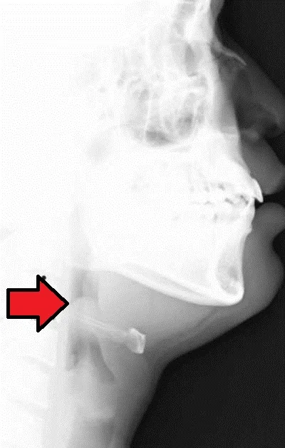
A lateral soft tissue neck radiograph may show the thumb sign of a swollen epiglottis, thickened aryepiglottic folds and ballooning of the hypopharynx. It has a real but narrow role: only in a stable child where the diagnosis is genuinely uncertain, only performed in the resuscitation area with a portable machine, only with the child sitting upright, and only with a clinician able to manage the airway present throughout. A normal film does not exclude the diagnosis.
Management
Immediate actions
- Call for help immediately - the most senior available anaesthetist, an ENT surgeon and a senior paediatrician. In most UK hospitals this is a pre-agreed emergency call, and it should be made on suspicion rather than after confirmation.
- Keep the child calm and upright on a parent's lap, with all painful and frightening procedures deferred
- Waft high-flow oxygen near the face without applying a mask
- Stay with the child and prepare, but do not use, airway equipment where the child can see it
- Prepare theatre, and have equipment for tracheostomy or needle cricothyroidotomy available as a rescue plan
Securing the airway
The definitive step is intubation under inhalational (gaseous) induction in theatre, performed by the most experienced anaesthetist available, with an ENT surgeon scrubbed and ready to perform a surgical airway if intubation fails.3,4
- Induction is inhalational with sevoflurane, with the child kept sitting up, and spontaneous ventilation maintained throughout
- Neuromuscular blockade is avoided at induction - if the airway is lost, the ability to keep breathing spontaneously is the only thing preventing complete obstruction
- An endotracheal tube half a size to a full size smaller than predicted for age is used, because the supraglottis is swollen
- A surgical airway - tracheostomy, or needle cricothyroidotomy as a temporising measure - is the backup plan and must be immediately available
- Blood cultures, a supraglottic swab and intravenous access are all obtained once the child is anaesthetised and intubated
Antibiotics and supportive care
- Intravenous ceftriaxone or cefotaxime is the standard empirical choice, covering Haemophilus influenzae, pneumococcus and group A streptococcus.5
- Add an antistaphylococcal agent such as flucloxacillin, or vancomycin where meticillin resistance is a concern, if staphylococcal infection is suspected
- Total course is usually 7-10 days, converting to oral once the child is extubated and improving
- Intravenous fluids while the child is nil by mouth
- Corticosteroids such as dexamethasone are often given to reduce supraglottic oedema before extubation, though the evidence for benefit is weak
- Management on paediatric intensive care while intubated, with sedation and adequate securing of the tube - accidental extubation in this setting is extremely dangerous
Most children are extubated within 24-48 hours, once fever has settled and a leak develops around the tube, indicating that the supraglottic swelling has resolved. Recovery thereafter is usually rapid and complete.
Complications
Complications fall into three groups: those of the obstruction itself, those of the associated bacteraemia, and those of the intervention needed to treat it. Recognising the second group matters, because a child who remains febrile after the airway is secured and antibiotics started should be examined for a metastatic focus rather than assumed to be responding slowly.
- Complete airway obstruction, respiratory arrest and hypoxic brain injury - the reason for everything above
- Epiglottic abscess, requiring surgical drainage
- Metastatic infection from the associated bacteraemia: pneumonia, meningitis, septic arthritis, osteomyelitis, cervical adenitis and pericarditis
- Sepsis and septic shock
- Post-obstructive pulmonary oedema after the obstruction is relieved, from the large negative intrathoracic pressures generated against a closed airway
- Complications of intubation - failed intubation, tube displacement, and subglottic stenosis after prolonged intubation
- Aspiration pneumonia from pooled secretions
Red flags
Prognosis
With the airway secured and appropriate antibiotics, recovery is the rule and long-term sequelae are uncommon. Mortality in hospital, where the airway is managed promptly, is well under 1%.6
The deaths that still occur happen before the child reaches a team able to secure the airway - during transfer, in a waiting room, or after an ill-judged attempt to look at the throat. This is the reason the management algorithm is built around not disturbing the child and calling for anaesthetic and ENT support before, rather than after, the diagnosis is confirmed.
At a population level, prognosis is now determined by immunisation. Hib vaccine uptake in the UK has historically been high and the disease has become rare, but the resurgence of vaccine-preventable illness where coverage falls means epiglottitis should stay on your list for any unimmunised child with fever, drooling and stridor - and the immunisation history should be documented in every such case.1
References
- UKHSA. Haemophilus influenzae type b (Hib): the Green Book, chapter 16. Available here
- UKHSA. Haemophilus influenzae type b (Hib): guidance, data and analysis. Available here
- Advanced Life Support Group. Advanced Paediatric Life Support: A Practical Approach to Emergencies. Available here
- Difficult Airway Society and Association of Paediatric Anaesthetists. Paediatric difficult airway guidelines. Available here
- BNF for Children. Ceftriaxone. Available here
- Guerra AM, Waseem M. Epiglottitis. StatPearls. 2023. Available here
This article is written for revision and education. It is not clinical guidance and must not be used to make decisions about the care of a patient. Always check current NICE guidance and local protocols.