
Adult Advanced Life Support
Key points
- First actions: confirm cardiac arrest (unresponsive, not breathing normally), call for help and the resuscitation team, and start CPR at 30 compressions to 2 breaths.
- Shockable rhythms: ventricular fibrillation and pulseless ventricular tachycardia - defibrillate immediately, then continue CPR.
- Non-shockable rhythms: asystole and pulseless electrical activity - no shock; give adrenaline as soon as access is obtained and continue CPR.
- Adrenaline: 1 mg IV every 3-5 minutes - immediately in non-shockable rhythms, after the third shock in shockable rhythms.
- Amiodarone: 300 mg IV after the third shock in refractory VF/pVT, with a further 150 mg after the fifth shock.
- Reversible causes: the 4Hs (hypoxia, hypovolaemia, hyper/hypokalaemia and metabolic, hypothermia) and 4Ts (tension pneumothorax, tamponade, toxins, thrombosis) - actively sought and treated during every cycle.
- Minimise interruptions: chest compressions should stop only for rhythm checks and defibrillation, kept as brief as possible.
- Post-ROSC care: controlled oxygenation, 12-lead ECG, treat the cause, and targeted temperature management for the comatose patient.
Introduction
Advanced life support (ALS) is the structured algorithm applied to a patient in cardiac arrest once basic life support has begun and a defibrillator or monitor is attached. It builds directly on basic life support - high-quality, minimally interrupted chest compressions remain the single intervention with the strongest evidence for survival, and everything else in ALS is added around that foundation.1
Survival from in-hospital and out-of-hospital cardiac arrest depends heavily on how quickly the chain of survival is completed: early recognition and call for help, early CPR, early defibrillation, and early post-resuscitation care. Every minute of delay to defibrillation in a shockable rhythm reduces the chance of survival by around 10%, which is why public-access defibrillators and rapid response teams exist - the algorithm only works if it is started promptly.
Survival to hospital discharge remains low overall (roughly 8-10% for out-of-hospital arrest in the UK, higher for in-hospital arrest and for an initial shockable rhythm), which is why so much emphasis is placed on the modifiable steps: starting CPR without delay, keeping interruptions to compressions as short as possible, and defibrillating a shockable rhythm as early as it is identified.
This article covers the adult in-hospital and out-of-hospital ALS algorithm as taught in the UK. Paediatric arrest, choking and basic life support sequences are covered separately, though the underlying principles overlap substantially.
Recognition and initial actions
Cardiac arrest is diagnosed clinically: the patient is unresponsive and not breathing normally (agonal gasps count as not breathing normally, and are a common source of confusion for bystanders and even clinical staff).
- Confirm unresponsiveness and absent normal breathing - assess for no more than 10 seconds
- Call for help - the resuscitation team (2222 in hospital, 999 in the community) and bring the defibrillator
- Start CPR at a ratio of 30 compressions to 2 breaths, compressions at a rate of 100-120/min and a depth of 5-6 cm, allowing full chest recoil between compressions
- Attach the defibrillator or monitor as soon as it arrives, and analyse the rhythm without stopping compressions for longer than necessary
- Assign roles - compressions, airway, defibrillator/drugs, team leader and documentation/timekeeping - if enough people are present
Rhythm classification and the algorithm
Once the rhythm is visible, cardiac arrest rhythms split into two groups that determine the entire subsequent pathway: shockable and non-shockable. Everything else - CPR, airway management, IV access, treating reversible causes - happens in both arms; the difference is whether a shock is delivered and when adrenaline is first given.
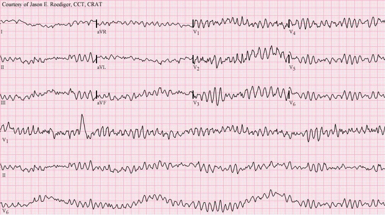
| Shockable | Non-shockable | |
|---|---|---|
| Rhythms | Ventricular fibrillation (VF), pulseless ventricular tachycardia (pVT) | Asystole, pulseless electrical activity (PEA) |
| First action | Defibrillate immediately | No shock - continue CPR |
| When to give adrenaline | After the 3rd shock, then every 3-5 min | As soon as IV/IO access is obtained, then every 3-5 min |
| Amiodarone | 300 mg after the 3rd shock; further 150 mg after the 5th shock | Not given |
| Rhythm/pulse check | Every 2 minutes, pausing compressions briefly | Every 2 minutes, pausing compressions briefly |
The shockable pathway (VF/pVT)
- Deliver a single shock (150-200 J biphasic, or per the manufacturer's guidance) with minimal interruption to compressions before and after
- Resume CPR immediately for 2 minutes without checking the rhythm or feeling for a pulse
- Re-check the rhythm - if still VF/pVT, deliver a second shock and resume CPR
- After the third shock, give adrenaline 1 mg IV and amiodarone 300 mg IV, then resume CPR
- Continue 2-minute cycles of CPR with a rhythm check between each; give adrenaline every 3-5 minutes (alternate cycles) and a further dose of amiodarone 150 mg after the fifth shock
The non-shockable pathway (PEA/asystole)
- Continue CPR in 2-minute cycles
- Give adrenaline 1 mg IV as soon as intravenous or intraosseous access is available, then every 3-5 minutes
- Re-check the rhythm every 2 minutes - if it becomes shockable, switch to the shockable pathway
- Actively search for and treat reversible causes throughout - this pathway has a lower spontaneous survival rate unless a reversible cause is identified and corrected
Airway and access during CPR
Airway management should not interrupt high-quality compressions. A basic airway with bag-valve-mask ventilation is often sufficient initially; a supraglottic airway (for example an i-gel) is commonly placed by the first appropriately trained provider, and once a supraglottic airway or tracheal tube is in place, compressions continue continuously at 100-120/min with asynchronous ventilations at 10/min.
- Intravenous access is first choice; if it cannot be obtained rapidly, use intraosseous access, which achieves comparable drug delivery
- Waveform capnography confirms tracheal tube placement, helps assess CPR quality, and a sudden rise can be the first sign of return of spontaneous circulation
- Tracheal intubation is reserved for those with the skill to do it without significant interruption to compressions - a prolonged, unsuccessful attempt is worse than a supraglottic airway placed quickly
Reversible causes: the 4Hs and 4Ts
Every cycle of CPR is an opportunity to look for and correct a reversible cause. In PEA and asystole in particular, survival is unlikely unless one of these is found and treated.
| Cause | Clues | Specific treatment |
|---|---|---|
| Hypoxia | Airway obstruction, respiratory failure, poor ventilation | Secure the airway, ventilate with high-flow oxygen |
| Hypovolaemia | Trauma, GI bleed, ruptured AAA, anaphylaxis | Rapid IV fluid and blood; control haemorrhage; treat anaphylaxis |
| Hyper/hypokalaemia, metabolic | Renal failure, dialysis, diabetic ketoacidosis, characteristic ECG changes | Calcium chloride, insulin/dextrose, sodium bicarbonate for hyperkalaemia; correct the underlying metabolic disturbance |
| Hypothermia | Cold water immersion, environmental exposure, elderly patient found collapsed | Active rewarming; continue resuscitation until core temperature is normal - 'not dead until warm and dead' |
| Tension pneumothorax | Trauma, asthma, absent breath sounds, tracheal deviation, raised JVP | Immediate needle decompression, then chest drain |
| Tamponade (cardiac) | Trauma, malignancy, post-cardiac surgery, raised JVP with muffled heart sounds | Pericardiocentesis |
| Toxins | Overdose, drug chart, collateral history, empty packets at scene | Specific antidote if available (for example naloxone, digoxin-specific antibody fragments); consider prolonged resuscitation with certain drugs |
| Thrombosis (coronary or pulmonary) | Preceding chest pain or breathlessness, risk factors for VTE, high clinical suspicion | Consider thrombolysis if massive PE is strongly suspected - if given, continue CPR for at least 60-90 minutes afterwards |
Drug doses at a glance
Beyond adrenaline and amiodarone, a handful of other drugs are used when a specific reversible cause is identified. Knowing the trigger for each is more useful than memorising the algorithm as a flat list.
| Drug | Dose | When |
|---|---|---|
| Adrenaline | 1 mg IV (10 mL of 1:10,000) | Every 3-5 min in all arrests - immediately in PEA/asystole, after the 3rd shock in VF/pVT |
| Amiodarone | 300 mg IV, then 150 mg | After the 3rd and 5th shocks in refractory VF/pVT |
| Lidocaine | 1 mg/kg IV | Alternative to amiodarone if unavailable, or as a second agent |
| Calcium chloride | 10 mL of 10% | Hyperkalaemia, hypocalcaemia, or calcium channel blocker overdose |
| Magnesium sulfate | 2 g IV | Torsades de pointes, or suspected hypomagnesaemia |
| Sodium bicarbonate | 50 mmol IV | Severe hyperkalaemia or tricyclic antidepressant overdose - not given routinely |
| Naloxone | 0.4-2 mg IV/IM | Known or suspected opioid-induced arrest, alongside standard ALS |
| Intravenous lipid emulsion | 1.5 mL/kg bolus | Local anaesthetic systemic toxicity |
Special circumstances
Several situations require a modification to the standard algorithm, either because the underlying physiology is different or because a specific intervention takes priority alongside standard CPR.
- Pregnancy - manual displacement of the uterus to the left (or a left lateral tilt) relieves aortocaval compression; involve obstetric and neonatal teams immediately, and perform a perimortem caesarean section within 5 minutes if there is no ROSC, both to improve maternal venous return and for fetal salvage
- Hyperkalaemia - calcium chloride to stabilise the myocardium, plus insulin/dextrose and salbutamol to shift potassium intracellularly; consider dialysis early with renal team input
- Hypothermic arrest - the myocardium is often unresponsive to drugs and defibrillation below 30°C; withhold or space out drug doses and shocks, and continue resuscitation during active rewarming since prognosis can be good even after prolonged arrest
- Local anaesthetic toxicity - IV lipid emulsion in addition to standard ALS
- Opioid overdose - naloxone alongside standard ALS and ventilation
Post-resuscitation care
Return of spontaneous circulation (ROSC) is the start of a new phase of management, not the end of the emergency. Post-resuscitation care aims to identify and treat the cause of arrest, limit secondary brain injury, and optimise the chance of a good neurological outcome.2
- Airway and breathing - secure the airway if not already done; titrate oxygen to a saturation of 94-98% (avoid hyperoxia, which is independently associated with worse outcomes) and maintain normocapnia
- Circulation - 12-lead ECG immediately; urgent coronary angiography if ST elevation or a strong suspicion of an acute coronary cause; maintain adequate blood pressure with fluids and vasopressors as needed
- Disability - manage seizures, maintain normoglycaemia, and avoid hyperthermia
- Targeted temperature management - actively prevent fever and maintain a constant target temperature (guidance has shifted towards normothermia, around 36°C, rather than deliberate hypothermia, but follow current local protocol) in the patient who remains comatose
- Identify and treat the cause - the same 4Hs and 4Ts framework applies to working out why the arrest happened
- Involve critical care early for ongoing organ support and neuroprognostication, which should not be attempted before at least 72 hours
When to stop resuscitation
The decision to stop is made by the team leader, in discussion with the team, and is based on the overall clinical picture rather than a fixed time limit. Resuscitation is generally discontinued when asystole persists despite 20 minutes of ALS with no reversible cause identified or correctable, though this is extended in hypothermia, drug toxicity, or where thrombolysis has been given.
A valid, accessible DNACPR (Do Not Attempt Cardiopulmonary Resuscitation) decision should be respected and CPR should not be started; if uncertainty exists about its validity at the time, current guidance is to begin resuscitation while the situation is clarified rather than to withhold it.
Red flags
References
- Resuscitation Council UK. Adult Advanced Life Support guidelines. Available here
- Resuscitation Council UK. Post-resuscitation care guidelines. Available here
- Soar J, Böttiger BW, Carli P et al. European Resuscitation Council Guidelines 2021: Adult advanced life support. Resuscitation. 2021. Available here
This article is written for revision and education. It is not clinical guidance and must not be used to make decisions about the care of a patient. Always check current NICE guidance and local protocols.