
Multiple Myeloma
Key points
- Multiple myeloma: a malignancy of clonal plasma cells within the bone marrow, producing a monoclonal immunoglobulin (paraprotein) and causing end-organ damage. Typically a disease of older adults (median age at diagnosis around 70).
- CRAB criteria: the defining end-organ damage: Calcium (hypercalcaemia), Renal impairment, Anaemia, Bone disease (lytic lesions/pathological fracture) - at least one is required alongside a clonal plasma cell population to diagnose active myeloma.
- Bone disease: caused by cytokine-driven (RANKL) osteoclast activation with suppressed osteoblast activity, producing lytic lesions, back pain and pathological fractures - not typically raised ALP, unlike most other bone pathology.
- Renal impairment: "myeloma kidney" - free light chains form obstructing casts in the distal tubule (cast nephropathy), compounded by hypercalcaemia, dehydration and hyperviscosity.
- Precursor states: MGUS (monoclonal gammopathy of undetermined significance) - a paraprotein without end-organ damage, with a small annual risk of progression - and smouldering myeloma, an intermediate, higher-risk state; neither is treated, only monitored.
- Diagnosis: serum/urine protein electrophoresis (paraprotein, Bence Jones protein), serum free light chains, skeletal survey or whole-body MRI/CT, and bone marrow biopsy (clonal plasma cells ≥10%).
- Management: combination therapy (commonly a proteasome inhibitor, immunomodulatory drug and dexamethasone), autologous stem cell transplant for eligible patients, and bisphosphonates for bone disease - myeloma remains incurable but treatable, with relapse expected.
- Emergencies: hypercalcaemia, spinal cord compression from a vertebral plasmacytoma or collapse, hyperviscosity syndrome, and acute kidney injury all need urgent recognition.
Introduction
Multiple myeloma is a malignancy of clonal plasma cells - terminally differentiated B lymphocytes that normally produce antibody - which accumulate within the bone marrow and secrete a monoclonal immunoglobulin or immunoglobulin fragment (paraprotein). It is the second commonest haematological malignancy after non-Hodgkin lymphoma, typically presenting in patients over 65.1
Myeloma sits at the malignant end of a spectrum of plasma cell disorders, with MGUS and smouldering myeloma as premalignant precursor states that are common, particularly with age, and require monitoring rather than treatment unless they progress.
Pathophysiology
Malignant plasma cells infiltrate the bone marrow and secrete cytokines (notably IL-6) that drive RANKL-mediated osteoclast activation while simultaneously suppressing osteoblast activity - the combination produces purely lytic bone destruction with little or no reparative response, which is why alkaline phosphatase is typically normal, unlike most other causes of bone pathology.
The paraprotein itself causes disease through several mechanisms: free light chains (kappa or lambda) are filtered by the kidney and can precipitate with Tamm-Horsfall protein to form obstructing casts in the distal tubule (myeloma cast nephropathy), and at high concentrations the paraprotein raises blood viscosity, impairing microcirculatory flow.
Clinical features and the CRAB criteria
Diagnosis of active (symptomatic) myeloma requires a clonal plasma cell population plus at least one feature of end-organ damage, remembered as CRAB:2
| Feature | Mechanism | Presentation |
|---|---|---|
| Calcium | Osteoclast-mediated bone resorption | Hypercalcaemia - "bones, stones, groans and psychiatric moans" |
| Renal impairment | Light chain cast nephropathy, hypercalcaemia, dehydration | Rising creatinine, may present as acute kidney injury |
| Anaemia | Marrow infiltration by plasma cells | Fatigue, pallor; normocytic, normochromic |
| Bone disease | RANKL-driven lytic lesions | Bone pain (classically back pain), pathological fracture, vertebral collapse |
Other common features include recurrent infections (from suppression of normal polyclonal immunoglobulin production - "immune paresis"), fatigue out of proportion to the anaemia, and symptoms of hyperviscosity (visual disturbance, headache, bleeding tendency, confusion) at high paraprotein levels.
Investigations
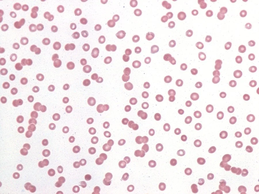
- FBC - normocytic, normochromic anaemia; the blood film shows rouleaux formation (red cells stacking like coins) from the raised paraprotein, and a very high ESR
- U&Es - renal impairment is common at presentation
- Corrected calcium - raised in a significant proportion at diagnosis
- Serum and urine protein electrophoresis - identifies the monoclonal band (paraprotein); urine electrophoresis detects Bence Jones protein (free light chains)
- Serum free light chain assay - particularly useful in light-chain-only or non-secretory myeloma, where whole immunoglobulin electrophoresis may be less sensitive
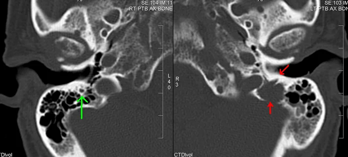
- Skeletal survey, or increasingly whole-body MRI or low-dose whole-body CT (more sensitive than plain films and now preferred where available), looking for lytic lesions - classically producing a "pepper-pot" skull appearance on plain radiographs where multiple lesions are present
- Bone marrow aspirate and trephine biopsy - confirms clonal plasma cells (≥10%) and allows cytogenetic risk stratification (e.g. t(4;14), del(17p) confer higher risk)
- Beta-2 microglobulin and albumin - used in the International Staging System (ISS), an important prognostic tool
Differential diagnosis and precursor states
| Condition | Paraprotein | Clonal plasma cells | CRAB features | Management |
|---|---|---|---|---|
| MGUS | <30 g/L | <10% | Absent | Monitor only; small annual risk (~1%) of progression to myeloma |
| Smouldering myeloma | ≥30 g/L and/or | 10-59% | Absent | Monitor closely; higher progression risk than MGUS |
| Symptomatic (active) myeloma | Usually present | ≥10% (or biopsy-proven plasmacytoma) | Present (or myeloma-defining biomarkers) | Treat |
Other differentials for a monoclonal paraprotein include Waldenstrom macroglobulinaemia (an IgM-secreting lymphoplasmacytic lymphoma causing hyperviscosity but typically without the lytic bone disease of myeloma) and amyloidosis (light chain deposition causing organ dysfunction, e.g. nephrotic syndrome, cardiomyopathy, without necessarily meeting CRAB criteria).
Management
- Combination systemic therapy - typically a proteasome inhibitor (e.g. bortezomib), an immunomodulatory drug (e.g. lenalidomide or thalidomide) and dexamethasone, with newer regimens increasingly incorporating anti-CD38 monoclonal antibodies (e.g. daratumumab)3
- Autologous stem cell transplant for fit, eligible patients following induction therapy, improving depth and duration of response, though not curative
- Bisphosphonates (e.g. zoledronic acid) for all patients with bone disease, reducing skeletal-related events and treating hypercalcaemia
- Radiotherapy for localised, painful bone lesions or impending fracture, and for cord compression
- Supportive care - analgesia, treatment of infections (with a low threshold given immune paresis), management of renal impairment and anaemia
- Relapse is expected - myeloma follows a relapsing-remitting course, and most patients receive multiple lines of treatment over time
MGUS and smouldering myeloma are not treated - only monitored - since treatment has not been shown to improve outcomes in these premalignant states and would expose patients to unnecessary toxicity.
Complications
- Renal failure - from cast nephropathy, hypercalcaemia and dehydration; may require dialysis
- Pathological fracture and spinal cord compression - from vertebral collapse or an epidural plasmacytoma; a same-day emergency
- Hypercalcaemia - can present acutely with confusion, dehydration and arrhythmia
- Hyperviscosity syndrome - visual disturbance, headache, bleeding, heart failure
- Recurrent infection - from immune paresis, a leading cause of death, particularly early in the disease course
- Amyloidosis - light chain deposition causing nephrotic syndrome, restrictive cardiomyopathy or peripheral neuropathy in a subset of patients
- Peripheral neuropathy - from the disease itself or as a side effect of treatment (notably bortezomib and thalidomide)
Red flags
Prognosis
Multiple myeloma remains incurable but increasingly treatable, with survival having improved substantially over the past two decades through proteasome inhibitors, immunomodulatory drugs and monoclonal antibodies. Median survival now extends well beyond 5-7 years for many patients, though this varies considerably with ISS stage and cytogenetic risk at diagnosis.1
The disease follows a relapsing-remitting course, with most patients cycling through multiple lines of treatment over years. MGUS carries an approximately 1% per year risk of progression to myeloma or a related malignancy and requires lifelong monitoring rather than treatment; smouldering myeloma carries a substantially higher progression risk and is monitored more closely, with early treatment now considered in selected high-risk cases.
References
- Kumar SK, Rajkumar V, Kyle RA et al. Multiple myeloma. Nat Rev Dis Primers. 2017. Available here
- Rajkumar SV, Dimopoulos MA, Palumbo A et al. International Myeloma Working Group updated criteria for the diagnosis of multiple myeloma. Lancet Oncol. 2014. Available here
- British Society for Haematology. Guidelines for the diagnosis and management of multiple myeloma. Available here
- Gabriel Caponetti, CC BY-SA 3.0, via Wikimedia Commons. Available here
- Dr Laughlin Dawes, radpod.org, CC BY 3.0, via Wikimedia Commons. Available here
- NICE. Myeloma: diagnosis and management (NG35). 2016 (updated 2018). Available here
This article is written for revision and education. It is not clinical guidance and must not be used to make decisions about the care of a patient. Always check current NICE guidance and local protocols.