
Fibroadenoma and Breast Cysts
Key points
- Fibroadenoma: a benign biphasic tumour of lobular stroma and epithelium. The commonest discrete breast lump in women under 30.
- Classic signs: a smooth, firm, non-tender, strikingly mobile lump - the breast mouse - typically 1 to 3 cm.
- Breast cyst: a fluid-filled space arising from involution of terminal duct lobular units. Typical of the perimenopausal years, 35 to 50.
- Cyst signs: a smooth, mobile, sometimes tender lump that may appear overnight, with a halo sign on mammography and posterior acoustic enhancement on ultrasound.
- ANDI: aberrations of normal development and involution - the framework that treats most benign breast disease as an exaggeration of physiology rather than a disease.
- Assessment: triple assessment for any discrete lump, with ultrasound first line under 40 and mammography plus ultrasound over 40.
- Managing a fibroadenoma: reassure and discharge if it is under 3 cm with concordant benign triple assessment. Excise if it is large, growing, symptomatic or discordant.
- The trap: a rapidly enlarging fibroadenoma-like lump, especially over 40, may be a phyllodes tumour, which requires wide local excision.
Introduction: the ANDI framework
Most benign breast conditions are not diseases at all but exaggerations of the normal cyclical processes of development, cyclical change and involution that the breast undergoes over a woman's reproductive life. The ANDI classification - Aberrations of Normal Development and Involution - organises them by the life stage in which they arise, and it is a considerably more useful mental model than a list of conditions.1
| Life stage | Normal process | Aberration | Disease |
|---|---|---|---|
| Early reproductive (15 to 25) | Lobular development | Fibroadenoma | Giant fibroadenoma, juvenile hypertrophy |
| Mature reproductive (25 to 40) | Cyclical hormonal change | Cyclical mastalgia, nodularity (fibrocystic change) | Incapacitating mastalgia |
| Involution (35 to 55) | Lobular involution and duct involution | Cysts, sclerosing adenosis, duct ectasia, nipple retraction | Periductal mastitis, periareolar abscess and fistula |
The practical value of this is that the age of the patient predicts the likely diagnosis. A discrete mobile lump in a woman of 22 is a fibroadenoma until proved otherwise; a discrete lump in a woman of 45 is a cyst until proved otherwise; and a discrete lump in a woman of 65 is a carcinoma until proved otherwise. Every one of them still requires triple assessment.
Fibroadenoma
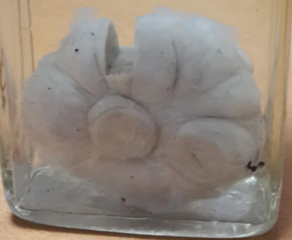
A fibroadenoma is a benign biphasic tumour containing both stromal and epithelial elements, arising from a single lobule. It is hormone-responsive, which is why it grows during pregnancy and with oestrogen exposure and typically regresses after the menopause.
Clinical features
- A smooth, firm, rubbery, well-defined lump, usually 1 to 3 cm
- Strikingly mobile within the breast tissue - it slips away from the examining fingers, which gives it the traditional name breast mouse
- Painless and non-tender, with no skin or nipple change and no lymphadenopathy
- Commonly in the upper outer quadrant, and multiple in around 10 to 20%
- Peak incidence between 15 and 35 years, and it is the commonest discrete breast lump in women under 30
- May fluctuate slightly with the menstrual cycle and enlarge during pregnancy and lactation
Types
- Simple fibroadenoma - the usual form, carrying no increased risk of subsequent breast cancer
- Complex fibroadenoma - contains cysts over 3 mm, sclerosing adenosis, epithelial calcification or papillary apocrine change. It carries a small increase in future breast cancer risk, roughly double the background rate, so these patients are not simply discharged.
- Giant fibroadenoma - greater than 5 cm, which may distort the breast and is usually excised
- Juvenile fibroadenoma - occurring in adolescence, growing rapidly and often reaching a large size, but histologically benign
Investigation and management
- Triple assessment, with ultrasound as the first-line imaging in women under 40. The typical appearance is a well-defined, oval, hypoechoic lesion that is wider than it is tall, with smooth margins and no posterior shadowing.
- Core biopsy where the lesion is over about 2.5 to 3 cm, where the patient is over 40, where the appearance is atypical, or where there is any discordance
- Reassure and discharge where the lump is under 3 cm, the imaging is characteristic and all three components of triple assessment agree it is benign. Explain the natural history: roughly a third regress, a third remain unchanged and a third enlarge, and most involute after the menopause.
- Excision or vacuum-assisted excision if the lesion is over 3 cm, is enlarging on surveillance, is symptomatic or causing distortion, is discordant on triple assessment, or if the patient wishes it removed after an informed discussion
- Safety-net advice - return if the lump grows, changes character, or any skin or nipple change develops
Breast cysts
Breast cysts are fluid-filled spaces formed when terminal duct lobular units involute and the acini distend, usually because the surrounding stroma involutes faster than the epithelium. They are extremely common, present in around 7% of women at some point and detectable on imaging in far more.
Clinical features
- A smooth, well-circumscribed, mobile lump, which may be tense and tender if it has filled quickly
- Sudden appearance - patients often describe a lump that was not there the day before, which is characteristic and reassuring rather than alarming
- Peak incidence between 35 and 50, in the perimenopausal years, and uncommon after the menopause unless the woman is on HRT
- Often multiple and bilateral, and may fluctuate with the cycle
- A halo sign on mammography - a thin lucent rim around a well-defined opacity
Ultrasound classification
| Type | Appearance | Management |
|---|---|---|
| Simple cyst | Anechoic, thin imperceptible wall, well circumscribed, with posterior acoustic enhancement | Benign. Aspirate only if symptomatic or the patient wishes it. No follow-up needed. |
| Complicated cyst | Low-level internal echoes or fluid-fluid levels, but no solid component or thick septation | Almost always benign. Aspiration to dryness, or short-interval ultrasound follow-up. |
| Complex cystic and solid lesion | Thick walls, thick septations, an intracystic mass, or a solid mural nodule | Requires core biopsy, since a small proportion are malignant - an intracystic papillary carcinoma or a necrotic tumour |
Management
- Triple assessment for any discrete lump, as always
- Aspiration of a symptomatic cyst, which is both diagnostic and therapeutic and gives immediate relief. Typical fluid is straw-coloured, green, brown or turbid - all of which are normal and do not need to be sent for cytology.
- Three findings after aspiration mandate further assessment: the aspirate is blood-stained, a residual mass remains after complete aspiration, or the cyst recurs rapidly or repeatedly at the same site. Any of these requires imaging and core biopsy.
- Reassure and discharge where the cyst resolves completely with a non-bloody aspirate and the rest of triple assessment is benign
- Simple cysts do not increase the risk of breast cancer and do not require surveillance
Other benign breast lumps
These appear in written papers as distractors, and each has a discriminating feature worth knowing.
| Condition | Typical patient | Distinguishing feature |
|---|---|---|
| Fibrocystic change (benign breast nodularity) | 25 to 45 | Diffuse, lumpy, tender breasts rather than a discrete lump, worse premenstrually and improving after a period. Bilateral. |
| Fat necrosis | Any age, often after trauma, surgery or radiotherapy, and in larger breasts | A firm, irregular, sometimes tethered lump that mimics carcinoma clinically and on imaging. There may be a history of trauma or a seatbelt injury, but often none. Core biopsy is usually needed to be sure. |
| Lipoma | Any age | Soft, smooth, mobile, fluctuant, slowly growing, with no skin or nipple change |
| Intraductal papilloma | 35 to 55 | Blood-stained or serous single-duct nipple discharge with or without a small subareolar lump. Requires excision (microdochectomy) because a papillary carcinoma cannot be reliably excluded on biopsy. |
| Duct ectasia | Perimenopausal, strongly associated with smoking | Dilated subareolar ducts producing thick, cheesy, green or creamy discharge from multiple ducts, with slit-like nipple retraction. Benign, but may become infected as periductal mastitis. |
| Sclerosing adenosis and radial scar | Any age | Usually detected on screening as a spiculated or distorted area that mimics carcinoma on mammography. Radial scars are excised or vacuum-excised because of an associated risk of adjacent malignancy. |
| Phyllodes tumour | 40 to 55 | Large, rapidly growing, fibroadenoma-like. Needs wide local excision. |
Mastalgia
Breast pain is the commonest breast symptom presenting to primary care, and it sits in the ANDI framework as an aberration of cyclical hormonal change rather than as a disease. It is worth covering here because the management is largely reassurance, and because the fear behind the consultation is usually cancer - which pain alone almost never indicates.
| Type | Features | Management |
|---|---|---|
| Cyclical mastalgia | Bilateral, diffuse, heaviest in the upper outer quadrants, worst in the luteal phase and relieved by menstruation. Peak age 30 to 40. | Reassurance, a well-fitting supportive bra, simple and topical analgesia, and a pain diary over two to three cycles |
| Non-cyclical mastalgia | Unrelated to the cycle, often unilateral and more localised. Occurs at any age including after the menopause. | Look for a specific cause - a cyst, duct ectasia, periductal mastitis, previous surgery or trauma - and treat it |
| Extramammary pain | Chest wall in origin - costochondritis, cervical or thoracic radiculopathy, shingles - or referred from the heart, gallbladder or lung | Reproduced by palpating the chest wall with the patient rolled onto the affected side, which moves the breast away. Treat the cause. |
- Take a full history including the drug chart - combined oral contraception, HRT, some antidepressants and antipsychotics all cause mastalgia
- Examine both breasts and axillae, and refer according to the standard criteria if a lump or other abnormality is found2
- Reassure explicitly that pain is not a symptom of cancer in the great majority of cases, which is the intervention that most patients actually came for
- A supportive, correctly fitted bra, worn during exercise and sometimes at night, has a genuine effect and is under-used
- Simple analgesia and topical NSAIDs are first-line pharmacological treatment
- Evening primrose oil is no longer recommended, since trials have not shown benefit over placebo
- Danazol and tamoxifen have a role in severe refractory cyclical mastalgia, prescribed by a specialist because of their side-effect profile3
Around 70 to 80% of women with cyclical mastalgia improve with reassurance and conservative measures within a few months, and a pain diary is often therapeutic in itself because it demonstrates the cyclical pattern and the periods of remission that the patient had not noticed.
Red flags
Prognosis and counselling
Both conditions are benign and neither shortens life. Simple fibroadenomas and simple cysts carry no meaningful increase in future breast cancer risk, and this is the single most useful thing to tell a patient, because the fear that prompted the referral is almost always about cancer rather than about the lump itself.
The exceptions are worth being precise about. Complex fibroadenomas, atypical ductal or lobular hyperplasia found in a biopsy, and radial scars all carry a modest increase in subsequent risk and warrant specialist follow-up rather than simple discharge. Proliferative changes without atypia carry a small increase; non-proliferative changes carry none.
A fibroadenoma left in situ follows a benign course, and there is no obligation to remove it. What matters is that the patient understands what has been excluded, knows what would prompt her to come back, and is not left with a vague reassurance she cannot act on. A specific instruction - return if it grows, changes shape, becomes fixed, or if the skin or nipple changes - is far more useful than telling her not to worry.
References
- Hughes LE, Mansel RE, Webster DJT. Aberrations of normal development and involution (ANDI): a new perspective on pathogenesis and nomenclature of benign breast disorders. The Lancet. 1987. Available here
- NICE Clinical Knowledge Summaries. Breast cancer - recognition and referral. Available here
- Association of Breast Surgery. Best practice guidelines. Available here
This article is written for revision and education. It is not clinical guidance and must not be used to make decisions about the care of a patient. Always check current NICE guidance and local protocols.